 Back
Cell Structure and Mitosis: Foundations of Cellular Biology
Study Guide - Smart Notes
 Tailored notes based on your materials, expanded with key definitions, examples, and context.
Cell Structure and the Basic Unit of Life
Cell Theory
Cells are the fundamental units of life, forming the basis for all living organisms. The cell theory states that all living things are composed of cells, and all cells arise from pre-existing cells. This concept underpins modern biology and explains the continuity of life.
All organisms are made of one or more cells.
The cell is the simplest collection of matter that can be alive.
All cells are related by descent from earlier cells.
Cells can differ substantially but share common features.
The Size Range of Cells
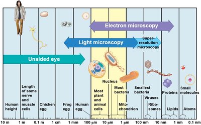
Cells vary greatly in size, from the smallest bacteria to large eukaryotic cells. The scale of cellular structures determines the methods used to study them, such as light or electron microscopy.
Prokaryotic cells are typically 0.1–5 μm in diameter.
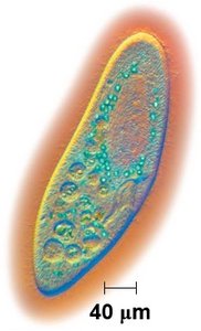
Eukaryotic cells are generally 10–100 μm in diameter.
Most plant and animal cells are visible with light microscopy, while smaller structures require electron microscopy.
Comparing Prokaryotic and Eukaryotic Cells
Basic Features of All Cells
Despite their diversity, all cells share several fundamental features:
Plasma membrane: Encloses the cell, controlling the movement of substances in and out.
Cytosol: Semifluid substance within the membrane.
Chromosomes: Carry genetic information (DNA).
Ribosomes: Synthesize proteins.
Prokaryotic Cells
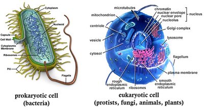
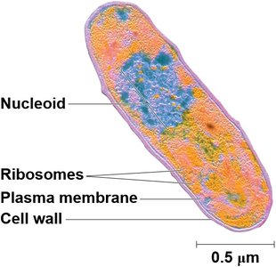
Prokaryotic cells, found in domains Bacteria and Archaea, lack a nucleus and membrane-bound organelles. Their DNA is located in a region called the nucleoid.
No nucleus; DNA is in the nucleoid.
No membrane-bound organelles.
Cytoplasm is bound by the plasma membrane.
Cell wall provides structural support.
Eukaryotic Cells
Eukaryotic cells are characterized by a true nucleus enclosed by a double membrane and numerous membrane-bound organelles. They are generally larger and more complex than prokaryotic cells.
Nucleus: Contains most of the cell's DNA.
Membrane-bound organelles: Specialized compartments (e.g., mitochondria, endoplasmic reticulum, Golgi apparatus).
Cytoplasm: Region between the plasma membrane and nucleus.
Prokaryotic Diversity and Ecological Roles
Abundance and Distribution
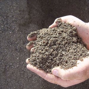
Prokaryotes are the most numerous and widespread organisms on Earth, inhabiting diverse environments from soil to the human gut.
More prokaryotic cells in a handful of soil than humans who have ever lived.
Prokaryotes outnumber eukaryotic cells in the human body.
Metabolic Diversity
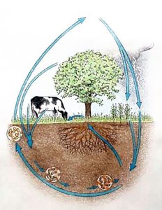
Prokaryotes exhibit remarkable metabolic diversity, serving as decomposers, recyclers, and symbionts in global nutrient cycles.
Decomposers: Break down dead organic matter.
Symbionts: Live in close association with other organisms (mutualists, commensalists, parasites).
Pathogens: Cause diseases in humans, animals, and plants.
Symbiotic Relationships of Prokaryotes
Types of Symbiosis
Prokaryotes form various symbiotic relationships with other organisms:
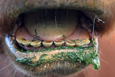
Mutualism: Both partners benefit (e.g., gut bacteria aiding digestion in herbivores).
Commensalism: One benefits, the other is unaffected.
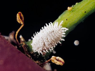
Parasitism: One benefits at the expense of the other (pathogens).
Metabolic Types in Prokaryotes
Sources of Carbon and Energy
Prokaryotes are classified by how they obtain carbon and energy:
Autotrophs: Use CO2 as a carbon source.
Heterotrophs: Require organic compounds for carbon.
Phototrophs: Use light as an energy source.
Chemotrophs: Obtain energy from chemical compounds.
Combinations include photoautotrophs, chemoautotrophs, photoheterotrophs, and chemoheterotrophs.
Examples of Metabolic Types
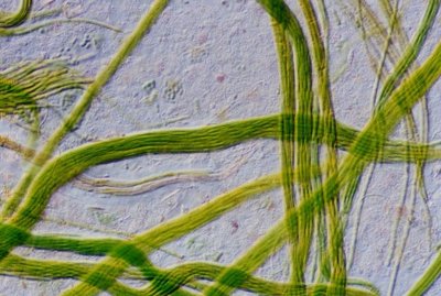
Photoautotrophs: Cyanobacteria (photosynthetic bacteria).
Chemoautotrophs: Oxidize inorganic substances (e.g., Methanococcus jannaschii).
Photoheterotrophs: Use light for energy but require organic carbon (e.g., Halobacterium salinarium).
Chemoheterotrophs: Obtain both energy and carbon from organic molecules (e.g., most bacteria, animals, fungi).
Nitrogen Metabolism
Some prokaryotes can fix atmospheric nitrogen (N2) into ammonia (NH3), making nitrogen available to other organisms. This process is essential for life on Earth.
Oxygen Relationships in Prokaryotes
Aerobic and Anaerobic Metabolism
Obligate aerobes: Require O2 for cellular respiration.
Facultative anaerobes: Use O2 if present, or switch to anaerobic metabolism.
Obligate anaerobes: Poisoned by O2; rely on anaerobic respiration or fermentation.
Early life was primarily anaerobic; the evolution of photosynthesis in cyanobacteria increased atmospheric oxygen.
Origin of Eukaryotes and Endosymbiosis
Endosymbiont Theory
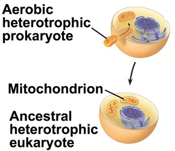
The endosymbiont theory proposes that mitochondria and chloroplasts originated as free-living prokaryotes that were engulfed by ancestral eukaryotic cells. This symbiotic relationship led to the evolution of complex eukaryotic cells.
Mitochondria evolved from aerobic heterotrophic prokaryotes (likely α-proteobacteria).
Chloroplasts evolved from photosynthetic prokaryotes (cyanobacteria).
Evidence: Both organelles have their own DNA, ribosomes, and double membranes; they replicate independently by binary fission.
Eukaryotic Cell Structure
Compartmentalization and Organelles
Eukaryotic cells contain membrane-bound organelles that compartmentalize cellular functions, increasing efficiency and complexity.
Nucleus: Stores genetic material.
Mitochondria: Site of cellular respiration and ATP production.
Chloroplasts: Site of photosynthesis in plants and algae.
Endoplasmic reticulum, Golgi apparatus, lysosomes: Involved in synthesis, modification, and transport of biomolecules.
The Cell Cycle and Mitosis
Roles of Cell Division
Cell division is essential for reproduction, growth, and repair in living organisms. In unicellular organisms, division produces new individuals; in multicellular organisms, it enables development, growth, and tissue repair.
Phases of the Cell Cycle
The cell cycle consists of interphase (G1, S, G2) and the mitotic (M) phase (mitosis and cytokinesis).
G1 phase: Cell growth.
S phase: DNA replication.
G2 phase: Preparation for mitosis.
M phase: Mitosis (nuclear division) and cytokinesis (cytoplasmic division).
Mitosis
Mitosis is the process by which a eukaryotic cell divides its nucleus and genetic material to produce two genetically identical diploid daughter cells. It is conventionally divided into five phases:
Prophase: Chromatin condenses into visible chromosomes; spindle apparatus forms.
Prometaphase: Nuclear envelope fragments; spindle fibers attach to kinetochores.
Metaphase: Chromosomes align at the metaphase plate.
Anaphase: Sister chromatids separate and move toward opposite poles.
Telophase: Nuclear envelopes reform; chromosomes decondense.
Cytokinesis follows, dividing the cytoplasm and completing cell division.
Chromosome Structure and DNA Packaging
In eukaryotes, DNA is packaged with proteins (histones) into chromatin, which condenses to form chromosomes during cell division. Chromosomes ensure accurate distribution of genetic material during mitosis.
Binary Fission and Evolution of Mitosis
Prokaryotes reproduce by binary fission, a simpler process than mitosis. Mitosis likely evolved from binary fission, with intermediate forms observed in some protists.
Summary Table: Prokaryotic vs. Eukaryotic Cells
Feature | Prokaryotic Cells | Eukaryotic Cells |
|---|---|---|
Nucleus | Absent (nucleoid region) | Present (membrane-bound) |
Membrane-bound organelles | Absent | Present |
Size | 0.1–5 μm | 10–100 μm |
Cell division | Binary fission | Mitosis and meiosis |
Examples | Bacteria, Archaea | Protists, fungi, plants, animals |
