 Back
BackCell Structure and Organisation: Study Notes for College Biology
Study Guide - Smart Notes
 Tailored notes based on your materials, expanded with key definitions, examples, and context.
Cell Theory
Foundations of Cell Theory
Cell theory is a fundamental concept in biology, describing the properties and significance of cells in living organisms. It was developed through the work of scientists such as Robert Hooke, Matthias Schleiden, and Theodor Schwann.
All living organisms are composed of one or more cells.
Cells are the basic functional unit in living organisms.
New cells arise from pre-existing cells.
Common features of all cells include a cell surface membrane, cytoplasm, DNA, and ribosomes.
Ultrastructure refers to the internal structures of the cell, visible under high magnification.
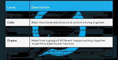
Levels of Organisation of Cells
Hierarchy in Multicellular Organisms
Cells in multicellular organisms are organised into increasingly complex structures, each with specific roles.
Specialised cells perform specific functions (e.g., red blood cells transport oxygen, xylem cells transport water in plants).
Tissues are groups of similar cells working together for a particular function (e.g., muscle tissue, epithelial tissue).
Organs are made from different tissues working together (e.g., heart, leaf).
Organ systems are groups of organs working together (e.g., circulatory system, digestive system).
Level | Description |
|---|---|
Cells | Basic functional and structural units in a living organism |
Organs | Made from a group of different tissues working together to perform a particular function |
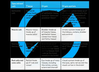
Examples of Specialised Cells and Organisation
Specialised cell | Tissue | Organ | Organ system |
|---|---|---|---|
Muscle cell | Muscle tissue | Bladder (muscle, epithelial, connective, fatty tissue) | Urinary system (kidneys, ureters, bladder, urethra) |
Rod cells and cone cells | Retina (rods and cones) | Eye (retina, cornea, sclera, choroid) | Visual system (eyes, optic nerves, visual cortex) |
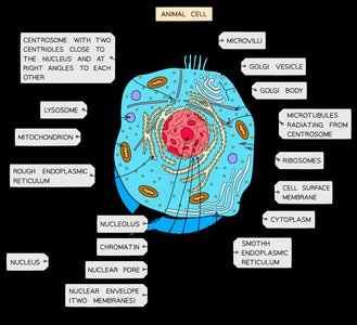
Eukaryotic Cells
General Structure
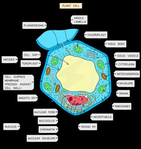
Eukaryotic cells are complex cells with membrane-bound organelles. They are generally larger than prokaryotic cells and include both animal and plant cells.
Diameter: 10–100 μm (eukaryotes) vs. 0.1–5 μm (prokaryotes)
Contain a nucleus and 80S ribosomes
Plant cells have a cellulose cell wall, large vacuole, and chloroplasts; animal cells have centrioles and sometimes microvilli
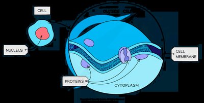
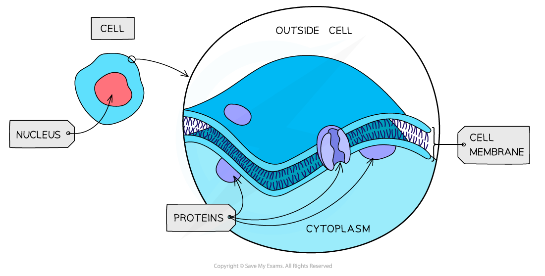
Cell Surface Membrane
The cell surface membrane (plasma membrane) controls the exchange of materials between the cell and its environment. It is partially permeable and composed of a phospholipid bilayer (~10 nm thick).
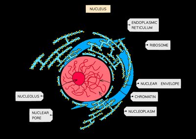
Nucleus
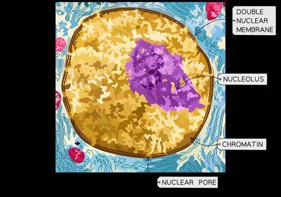
The nucleus is the control center of the cell, containing genetic material and surrounded by a double membrane (nuclear envelope) with pores for molecular exchange.
Contains chromatin (DNA + histones)
Has a nucleolus for ribosome production
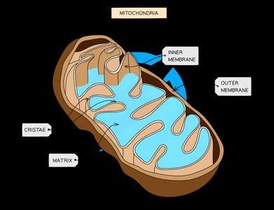
Mitochondria
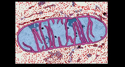
Mitochondria are the site of aerobic respiration, producing ATP. They have a double membrane, with the inner membrane folded into cristae and a matrix containing enzymes, mitochondrial DNA, and ribosomes.
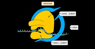
Ribosomes
Ribosomes are the site of protein synthesis (translation). Eukaryotic cells have 80S ribosomes, while prokaryotes, mitochondria, and chloroplasts have 70S ribosomes. Ribosomes are made of rRNA and proteins.
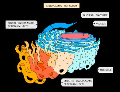
Endoplasmic Reticulum (ER)
The ER is a network of membranes involved in synthesis and transport. There are two types:
Rough ER (RER): Studded with ribosomes; processes and folds proteins.
Smooth ER (SER): Lacks ribosomes; synthesizes and processes lipids, carbohydrates, and steroids.
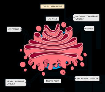
Golgi Apparatus
The Golgi apparatus modifies, sorts, and packages proteins and lipids for secretion or delivery to other organelles. It consists of stacked, flattened membrane sacs (cisternae).
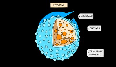
Lysosomes
Lysosomes are vesicles containing hydrolytic enzymes for breaking down waste, old organelles, and pathogens. They play a role in apoptosis (programmed cell death).
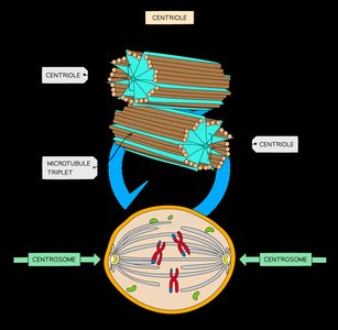
Centrioles
Centrioles are cylindrical structures made of microtubules, involved in organizing spindle fibers during cell division. They are found in animal cells but not in plants or fungi.
The Rough Endoplasmic Reticulum & Golgi Apparatus
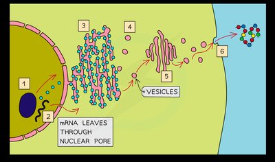
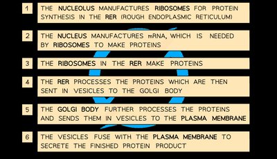
Protein Synthesis and Secretion Pathway
Several organelles work together to produce and secrete proteins, especially extracellular enzymes:
Nucleus: Transcription of DNA to mRNA
Ribosomes: Translation of mRNA to protein (free ribosomes for cytoplasmic proteins; RER-bound for secreted/membrane proteins)
RER: Folding and processing of proteins
Golgi apparatus: Further modification and packaging into vesicles
Cell surface membrane: Vesicles fuse to secrete proteins
Prokaryotic Cells
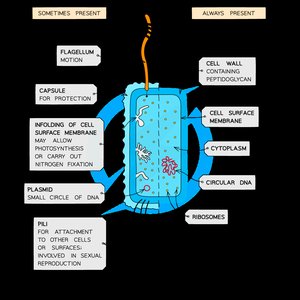
Structure and Features
Prokaryotic cells (e.g., bacteria) are simpler and smaller than eukaryotic cells. They lack membrane-bound organelles and a true nucleus.
Cytoplasm without membrane-bound organelles
70S ribosomes
Single circular DNA (not associated with proteins)
Cell wall made of murein (peptidoglycan)
May have plasmids, capsule, flagella, pili, and mesosomes
Prokaryotes vs. Eukaryotes: Comparison Table
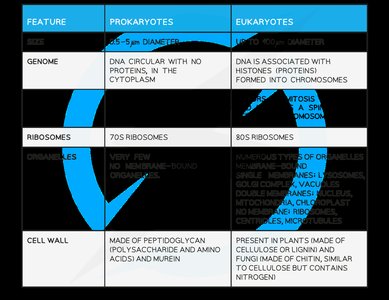
Feature | Prokaryotes | Eukaryotes |
|---|---|---|
Genome | DNA circular, no proteins, in cytoplasm | DNA with histones, in nucleus |
Ribosomes | 70S | 80S |
Organelles | Very few, no membrane-bound | Numerous, membrane-bound |
Cell wall | Peptidoglycan (murein) | Cellulose (plants), chitin (fungi), or absent (animals) |
Additional Prokaryotic Structures
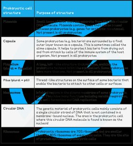
Prokaryotic cell structure | Purpose of structure |
|---|---|
Plasmid | Small, circular DNA; may carry genes for antibiotic resistance |
Capsule | Protects from desiccation and immune attack |
Pilus | Attachment to surfaces or other cells; involved in conjugation |
Flagellum | Movement |
Circular DNA | Genetic material, not in a nucleus |
Electron Microscopy of Animal Cells
Types of Electron Microscopes
Electron microscopes provide high-resolution images of cell ultrastructure. There are two main types:
Transmission Electron Microscope (TEM): Electrons pass through thin specimens, producing high-resolution 2D images of internal structures.
Scanning Electron Microscope (SEM): Electrons scan the surface, producing 3D images of specimen surfaces.
Microscopy: Magnification & Resolution
Magnification
Magnification is the ratio of the image size to the actual size of the specimen. In light microscopy, total magnification is calculated as:
Resolution
Resolution is the ability to distinguish two points as separate. Light microscopes have a maximum resolution of 200 nm, while electron microscopes can resolve structures as small as 0.5 nm due to the shorter wavelength of electrons.
Comparing Light and Electron Microscopes
Light microscopes: lower resolution, can view living specimens, suitable for whole cells and tissues.
Electron microscopes: higher resolution, only dead specimens, suitable for organelles, viruses, and DNA.
Staining Specimens
Purpose and Types of Stains
Staining enhances contrast in microscopic images, making cell structures more visible. Different stains are used for different cell components:
Haemotoxylin: Nuclei (purple, brown, blue)
Methylene blue: Animal cell nuclei (blue)
Acetocarmine: Chromosomes (red)
Iodine: Starch (blue-black)
Toluidine blue: DNA and RNA (blue)
Phloroglucinol: Lignin (red/pink)
Core Practical 5 - Light Microscopy
Microscope Use and Slide Preparation
Light microscopes are essential for studying cells and tissues. Proper slide preparation and staining are crucial for clear observation.
Start with low power objective lens to locate specimen
Use stains to enhance visibility
Thin tissue sections are necessary for light penetration
Prevent dehydration by adding water under coverslip
Drawing Cells
Biological drawings should be clear, accurate, and labeled, following conventions such as no shading, proper proportions, and including magnification.
Measuring Microscopic Images
Magnification can also be calculated as:
Eyepiece graticules and stage micrometers are used to measure objects under the microscope, requiring calibration for each objective lens.
