 Back
BackCell Structure, Function, and Microscopy: Foundations of Life
Study Guide - Smart Notes
 Tailored notes based on your materials, expanded with key definitions, examples, and context.
Cells as the Basis of Life
Prokaryotic and Eukaryotic Cells
Cells are the fundamental units of life, classified into two main types: prokaryotic and eukaryotic. Prokaryotic cells are simple, single-celled organisms lacking a true nucleus and membrane-bound organelles, while eukaryotic cells are more complex, containing a nucleus and various organelles.
Prokaryotic Cells: Found in bacteria and archaea; DNA is free-floating in the cytoplasm.
Eukaryotic Cells: Found in animals, plants, fungi, and protists; DNA is enclosed within a nucleus.
Similarities: Both types have DNA, a cell membrane, and cytoplasm.
Differences: Eukaryotes have a nucleus and membrane-bound organelles; prokaryotes do not.
Microscopy and Cell Structure
Microscope Technologies: Magnification and Resolution
Microscopes are essential tools for studying cells. Two key properties are magnification (enlarging the image) and resolution (clarity and ability to distinguish details).
Light Microscopes: Use visible light and glass lenses; magnification up to x1500; resolution ~200nm; can view living and stained specimens.
Electron Microscopes: Use electron beams; higher magnification (up to x1,500,000) and resolution (~2nm); TEM produces 2D images, SEM produces 3D images; only non-living specimens can be viewed.
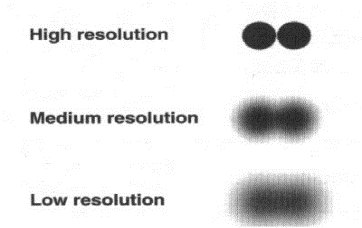
Resolution determines how well two points can be distinguished as separate. Higher resolution provides clearer, more detailed images.
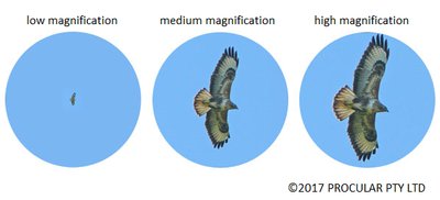
Magnification increases the size of the specimen's image, but the field of view decreases as magnification increases.
Cell Organelles: Structure and Function
Eukaryotic cells contain specialized organelles, each with distinct structures and functions.
Nucleus
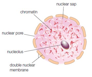
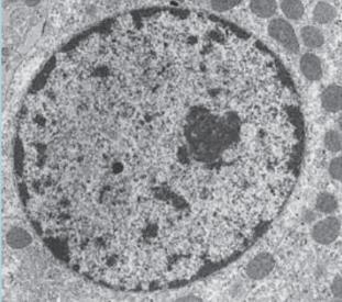
The nucleus is the control center of the cell, storing DNA and regulating cellular activities. It is surrounded by a double nuclear membrane with pores for material exchange.
Nucleolus: Produces ribosomes.
Nuclear pores: Allow passage of substances between nucleus and cytoplasm.
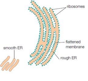
Endoplasmic Reticulum (ER)
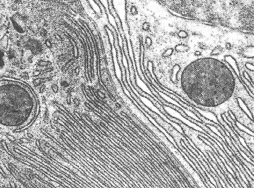
The endoplasmic reticulum is a network of membranes involved in protein and lipid synthesis.
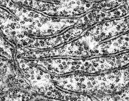
Rough ER: Studded with ribosomes; processes proteins.
Smooth ER: Lacks ribosomes; synthesizes lipids.
Ribosomes
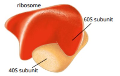
Ribosomes are the sites of protein synthesis, found free in the cytoplasm or attached to the rough ER.
Composed of two subunits (large and small).
Translate mRNA into polypeptides.
Golgi Apparatus
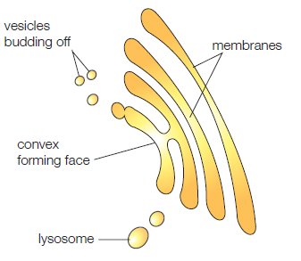
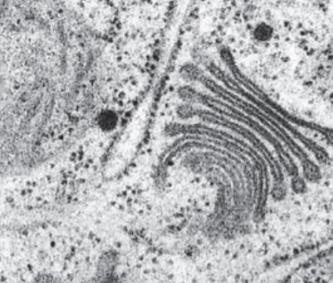
The Golgi apparatus processes, packages, and sorts cell products, especially proteins, into vesicles for transport.
Consists of stacked, flattened membranes.
Vesicles bud off for transport.
Lysosomes
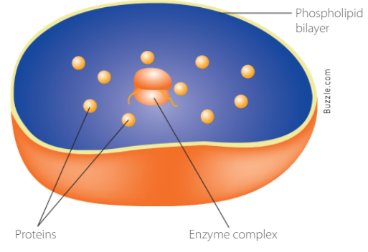
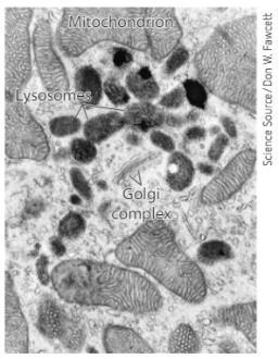
Lysosomes are membrane-bound vesicles containing digestive enzymes, responsible for breaking down waste and recycling materials.
Mitochondria
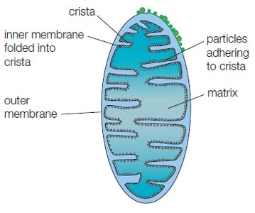
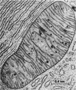
Mitochondria are the site of aerobic respiration, producing ATP energy. They have a double membrane, with the inner membrane highly folded (cristae) to increase surface area for reactions.
Contain their own DNA.
ATP production via cellular respiration.
Vacuole
Vacuoles are membrane-bound, fluid-filled vesicles for storage and maintaining cell shape, especially large in plant cells.
Chloroplasts
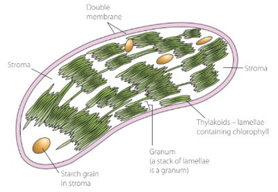
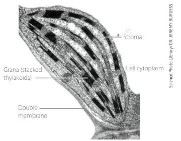
Chloroplasts are found in plant cells, containing chlorophyll for photosynthesis. They have a double membrane and stacks of thylakoids.
Site of photosynthesis: conversion of light energy to chemical energy (glucose).
Cell Wall
The cell wall is an external structure found in plant cells, providing support and protection.
Cell Membrane
The cell membrane is a semi-permeable phospholipid bilayer that controls the movement of substances into and out of the cell.
Contains embedded proteins for transport and communication.
Maintains homeostasis.
Fluid Mosaic Model of the Cell Membrane
Structure and Function
The fluid mosaic model describes the cell membrane as a dynamic structure with a phospholipid bilayer and embedded proteins.
Phospholipids: Form a bilayer; hydrophilic heads face outward, hydrophobic tails inward.
Proteins: Integral and peripheral proteins facilitate transport and communication.
Cholesterol: Provides stability and fluidity.
Carbohydrates: Glycoproteins and glycolipids aid in cell recognition and adhesion.
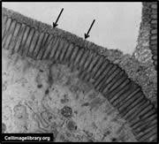
Movement of Materials Across Membranes
Passive and Active Transport
Cells exchange materials with their environment through various mechanisms:
Passive Transport: No energy required; includes diffusion, osmosis, and facilitated diffusion.
Active Transport: Requires energy (ATP); moves substances against concentration gradients.
Diffusion
Movement of molecules from high to low concentration until equilibrium is reached.
Osmosis
Movement of water across a selectively permeable membrane from high to low water concentration, often through aquaporins.
Facilitated Diffusion
Transport of large or charged molecules via channel or carrier proteins.
Active Transport, Endocytosis, and Exocytosis
Active Transport: Uses carrier proteins and ATP to move substances from low to high concentration.
Endocytosis: Cell engulfs particles (phagocytosis for solids, pinocytosis for liquids).
Exocytosis: Vesicles fuse with membrane to release contents outside the cell.
Surface Area to Volume Ratio
The efficiency of material exchange depends on the cell's surface area to volume ratio (SA:V). Smaller cells have a larger SA:V, allowing faster exchange and optimal functioning.
Cell Requirements: Organic and Inorganic Compounds
Organic Compounds
Carbohydrates: Energy sources and structural components (e.g., glucose, starch, cellulose).
Lipids: Energy storage and membrane structure (triglycerides).
Proteins: Structural, enzymatic, and hormonal functions; built from amino acids.
Nucleic Acids: DNA and RNA; store and transmit genetic information.
Inorganic Compounds and Waste Removal
Cells require gases (O2, CO2), ions, and water.
Waste products must be removed to prevent interference with cell function (excretion).
Lysosomes and exocytosis are involved in waste removal.
Enzymes and Metabolism
Enzyme Structure and Function
Enzymes are proteins that act as biological catalysts, speeding up chemical reactions by lowering activation energy.
Each enzyme has a specific shape and active site for substrate binding.
Enzyme-substrate complex forms during catalysis.
Lock and Key vs. Induced Fit Models
Lock and Key: Substrate fits exactly into rigid active site.
Induced Fit: Active site changes shape to fit substrate.
Coenzymes
Non-protein molecules (e.g., vitamins, metal ions) required for enzyme function.
Factors Affecting Enzyme Activity
Temperature: Optimum at 37°C; high temperatures denature enzymes.
pH: Each enzyme has an optimal pH; extreme pH denatures enzymes.
Substrate Concentration: Increased concentration increases rate until saturation point.
Summary Table: Cell Organelles and Functions
Organelle | Structure | Function |
|---|---|---|
Nucleus | Double membrane, pores, nucleolus | Stores DNA, controls cell activities |
ER (Rough/Smooth) | Network of membranes, ribosomes (rough) | Protein and lipid synthesis |
Ribosomes | Two subunits | Protein synthesis |
Golgi Apparatus | Stacked membranes | Processing and packaging |
Lysosomes | Membrane-bound vesicles | Digestive enzymes, waste removal |
Mitochondria | Double membrane, cristae | ATP production |
Vacuole | Membrane-bound, fluid-filled | Storage, turgor pressure |
Chloroplast | Double membrane, thylakoids | Photosynthesis |
Cell Wall | External, rigid | Support, protection |
Cell Membrane | Phospholipid bilayer | Selective permeability |
Equations and Calculations
Total Magnification: $\text{Total Magnification} = \text{ocular lens} \times \text{objective lens}$
Size of Cell: $\text{Size of cell} = \frac{\text{FOV}}{\text{number of cells across diameter}}$
Surface Area of Cube: $\text{SA} = \text{number of faces} \times \text{height} \times \text{length}$
Volume of Cube: $\text{Volume} = \text{height} \times \text{length} \times \text{width}$
Example: Plant Cell Structure
Plant cells contain all major organelles, including cell wall, chloroplasts, and a large central vacuole.
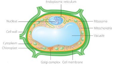
Example: Chloroplast Structure
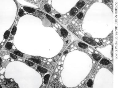
Example: Mitochondrion Structure
Example: Cell Membrane Structure
Example: Golgi Apparatus Structure
Example: Endoplasmic Reticulum Structure
Example: Nucleus Structure
Example: Ribosome Structure
Example: Plant Cell with Organelles
Example: Cell Wall Structure
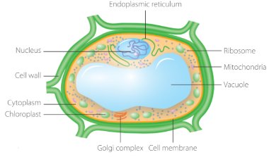
Example: Fluid Mosaic Model
Example: Mitochondrion Structure
Example: Chloroplast Structure
Example: Plant Cell Structure
Example: Cell Wall Structure
Example: Fluid Mosaic Model
Example: Mitochondrion Structure
Example: Chloroplast Structure
Example: Plant Cell Structure
Example: Cell Wall Structure
Example: Fluid Mosaic Model
Example: Mitochondrion Structure
Example: Chloroplast Structure
Example: Plant Cell Structure
Example: Cell Wall Structure
Example: Fluid Mosaic Model
Example: Mitochondrion Structure
Example: Chloroplast Structure
Example: Plant Cell Structure
Example: Cell Wall Structure
Example: Fluid Mosaic Model
Example: Mitochondrion Structure
Example: Chloroplast Structure
Example: Plant Cell Structure
Example: Cell Wall Structure
Example: Fluid Mosaic Model
Example: Mitochondrion Structure
Example: Chloroplast Structure
Example: Plant Cell Structure
Example: Cell Wall Structure
Example: Fluid Mosaic Model
Example: Mitochondrion Structure
Example: Chloroplast Structure
Example: Plant Cell Structure
Example: Cell Wall Structure
Example: Fluid Mosaic Model
Example: Mitochondrion Structure
Example: Chloroplast Structure
Example: Plant Cell Structure
Example: Cell Wall Structure
Example: Fluid Mosaic Model
Example: Mitochondrion Structure
Example: Chloroplast Structure
Example: Plant Cell Structure
Example: Cell Wall Structure
Example: Fluid Mosaic Model
Example: Mitochondrion Structure
Example: Chloroplast Structure
Example: Plant Cell Structure
Example: Cell Wall Structure
Example: Fluid Mosaic Model
Example: Mitochondrion Structure
Example: Chloroplast Structure
Example: Plant Cell Structure
Example: Cell Wall Structure
Example: Fluid Mosaic Model
Example: Mitochondrion Structure
Example: Chloroplast Structure
Example: Plant Cell Structure
Example: Cell Wall Structure
Example: Fluid Mosaic Model
Example: Mitochondrion Structure
Example: Chloroplast Structure
Example: Plant Cell Structure
Example: Cell Wall Structure
Example: Fluid Mosaic Model
Example: Mitochondrion Structure
Example: Chloroplast Structure
Example: Plant Cell Structure
Example: Cell Wall Structure
Example: Fluid Mosaic Model
Example: Mitochondrion Structure
Example: Chloroplast Structure
Example: Plant Cell Structure
Example: Cell Wall Structure
Example: Fluid Mosaic Model
Example: Mitochondrion Structure
Example: Chloroplast Structure
Example: Plant Cell Structure
Example: Cell Wall Structure
Example: Fluid Mosaic Model
Example: Mitochondrion Structure
Example: Chloroplast Structure
Example: Plant Cell Structure
Example: Cell Wall Structure
Example: Fluid Mosaic Model
Example: Mitochondrion Structure
Example: Chloroplast Structure
Example: Plant Cell Structure
Example: Cell Wall Structure
Example: Fluid Mosaic Model
Example: Mitochondrion Structure
Example: Chloroplast Structure
Example: Plant Cell Structure
Example: Cell Wall Structure
Example: Fluid Mosaic Model
Example: Mitochondrion Structure
Example: Chloroplast Structure
Example: Plant Cell Structure
Example: Cell Wall Structure
Example: Fluid Mosaic Model
Example: Mitochondrion Structure
Example: Chloroplast Structure
Example: Plant Cell Structure
Example: Cell Wall Structure
Example: Fluid Mosaic Model
Example: Mitochondrion Structure
Example: Chloroplast Structure
Example: Plant Cell Structure
Example: Cell Wall Structure
Example: Fluid Mosaic Model
Example: Mitochondrion Structure
Example: Chloroplast Structure
Example: Plant Cell Structure
Example: Cell Wall Structure
Example: Fluid Mosaic Model
Example: Mitochondrion Structure
Example: Chloroplast Structure
Example: Plant Cell Structure
Example: Cell Wall Structure
Example: Fluid Mosaic Model
Example: Mitochondrion Structure
Example: Chloroplast Structure
Example: Plant Cell Structure
Example: Cell Wall Structure
Example: Fluid Mosaic Model
Example: Mitochondrion Structure
Example: Chloroplast Structure
Example: Plant Cell Structure
Example: Cell Wall Structure
Example: Fluid Mosaic Model
Example: Mitochondrion Structure
Example: Chloroplast Structure
Example: Plant Cell Structure
Example: Cell Wall Structure
Example: Fluid Mosaic Model
Example: Mitochondrion Structure
Example: Chloroplast Structure
Example: Plant Cell Structure
Example: Cell Wall Structure
Example: Fluid Mosaic Model
Example: Mitochondrion Structure
Example: Chloroplast Structure
Example: Plant Cell Structure
Example: Cell Wall Structure
Example: Fluid Mosaic Model
Example: Mitochondrion Structure
Example: Chloroplast Structure
Example: Plant Cell Structure
Example: Cell Wall Structure
Example: Fluid Mosaic Model
Example: Mitochondrion Structure
Example: Chloroplast Structure
Example: Plant Cell Structure
Example: Cell Wall Structure
Example: Fluid Mosaic Model
Example: Mitochondrion Structure
Example: Chloroplast Structure
Example: Plant Cell Structure
Example: Cell Wall Structure
