 Back
Cell Structure, Function, and Microscopy: General Biology Study Guide
Study Guide - Smart Notes
 Tailored notes based on your materials, expanded with key definitions, examples, and context.
Q1. Describe differences between light and electron microscopes, and how each is used to visualize the structure of cells.
Background
Topic: Microscopy and Cell Visualization
This question tests your understanding of the types of microscopes used in biology and their applications for visualizing cell structures.
Key Terms:
Light Microscope: Uses visible light to illuminate specimens; suitable for viewing living cells and larger cell structures.
Electron Microscope: Uses beams of electrons for much higher resolution; suitable for viewing very small structures like organelles and ribosomes.
Step-by-Step Guidance
Identify the main differences in how light and electron microscopes work (light vs. electron beams).
Consider what structures are visible with each type: light microscopes can see cells and some organelles, electron microscopes can see much smaller details.
Think about the advantages and limitations of each: light microscopes allow observation of living cells, electron microscopes provide greater detail but require dead specimens.
Review examples of cell structures that are best visualized with each microscope.
Try solving on your own before revealing the answer!
Final Answer:
Light microscopes use visible light and are best for viewing living cells and larger structures (e.g., nucleus, cell membrane). Electron microscopes use electron beams and can visualize much smaller structures (e.g., ribosomes, mitochondria) with higher resolution. Transmission electron microscopes (TEM) show internal details, while scanning electron microscopes (SEM) show surface textures.
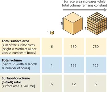
Q2. Explain how the surface area-to-volume ratio affects cell size and the efficiency of cellular processes, and why cells must remain small.
Background
Topic: Cell Size and Surface Area-to-Volume Ratio
This question tests your understanding of why cells are small and how their size impacts their function.
Key Terms and Formulas:
Surface Area: The total area of the cell's membrane.
Volume: The space inside the cell.
Surface Area-to-Volume Ratio:
Step-by-Step Guidance
Recall that as a cell grows, its volume increases faster than its surface area.
Understand that a high surface area-to-volume ratio is important for efficient transport of materials in and out of the cell.
Use the formula for surface area and volume to compare how the ratio changes as cell size increases.
Think about the consequences for cells if the ratio becomes too low.
Review why cells must divide or stay small to maintain efficient function.
Try solving on your own before revealing the answer!
Final Answer:
As cells increase in size, their surface area-to-volume ratio decreases, making it harder for materials to move efficiently across the membrane. Cells must remain small or divide to maintain a high ratio, which is essential for efficient diffusion and cellular communication.
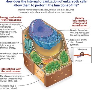
Q3. Describe the key structures and functions of prokaryotic and eukaryotic cells.
Background
Topic: Cell Types and Structures
This question tests your knowledge of the differences and similarities between prokaryotic and eukaryotic cells.
Key Terms:
Prokaryotic Cell: Lacks membrane-bound organelles; DNA in nucleoid; has cell wall, ribosomes, plasma membrane.
Eukaryotic Cell: Has membrane-bound organelles (nucleus, mitochondria, etc.); DNA in nucleus; may have cell wall (plants), ribosomes, plasma membrane.
Step-by-Step Guidance
List the basic features all cells share: plasma membrane, ribosomes, cytoplasm, DNA.
Identify unique features of prokaryotes: cell wall made of peptidoglycan, circular DNA, no nucleus.
Identify unique features of eukaryotes: nucleus, organelles, linear DNA.
Compare the functions of these structures in both cell types.
Try solving on your own before revealing the answer!
Final Answer:
Both prokaryotic and eukaryotic cells have plasma membranes, ribosomes, cytoplasm, and DNA. Prokaryotes lack membrane-bound organelles and have circular DNA, while eukaryotes have a nucleus and other organelles, with linear DNA. These differences impact cell function and complexity.
Q4. Describe how membrane-bound organelles help compartmentalize cellular functions.
Background
Topic: Cellular Compartmentalization
This question tests your understanding of how organelles allow cells to perform specialized functions efficiently.
Key Terms:
Organelle: Specialized structure within a cell, surrounded by a membrane.
Compartmentalization: Separation of cellular processes into different areas.
Step-by-Step Guidance
Recall that organelles such as the nucleus, mitochondria, and ER are surrounded by membranes.
Understand that these membranes create separate environments for different biochemical reactions.
Think about how compartmentalization increases efficiency and prevents interference between processes.
Review examples of organelles and their specialized functions.
Try solving on your own before revealing the answer!
Final Answer:
Membrane-bound organelles compartmentalize cellular functions, allowing different metabolic processes to occur simultaneously and efficiently. This separation prevents interference and increases the cell's ability to specialize and adapt.
Q5. Explain the structure and role of the cytoskeleton in supporting cell shape, enabling movement, and organizing cell structures.
Background
Topic: Cytoskeleton Structure and Function
This question tests your understanding of the cytoskeleton's components and their roles in cell function.
Key Terms:
Cytoskeleton: Network of protein filaments (microtubules, actin filaments, intermediate filaments).
Microtubules: Provide structural support, move organelles, involved in cell division.
Actin Filaments: Enable cell movement, muscle contraction, and cell division.
Intermediate Filaments: Maintain cell shape and anchor organelles.
Step-by-Step Guidance
Identify the three main components of the cytoskeleton.
Describe the role of each component in maintaining cell shape and movement.
Consider what would happen if one or more components were missing.
Review examples of cells that rely heavily on the cytoskeleton.
Try solving on your own before revealing the answer!
Final Answer:
The cytoskeleton consists of microtubules, actin filaments, and intermediate filaments. It supports cell shape, enables movement, and organizes cell structures. Without these filaments, cells would lose shape, have impaired movement, and fail to transport materials efficiently.
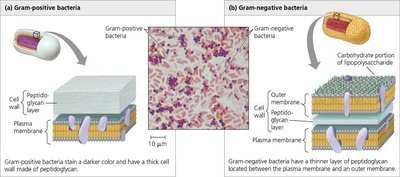
Q6. Using the figure, describe the structural differences in gram positive vs gram negative bacteria.
Background
Topic: Bacterial Cell Wall Structure
This question tests your ability to distinguish between gram-positive and gram-negative bacteria based on cell wall structure.
Key Terms:
Gram-positive: Thick peptidoglycan layer, no outer membrane.
Gram-negative: Thin peptidoglycan layer, outer membrane present.
Step-by-Step Guidance
Examine the diagram showing the cell wall structure of both types.
Identify the thickness of the peptidoglycan layer in each.
Note the presence or absence of an outer membrane.
Consider how these differences affect staining and antibiotic susceptibility.
Try solving on your own before revealing the answer!
Final Answer:
Gram-positive bacteria have a thick peptidoglycan layer and no outer membrane, making them stain darker and more susceptible to antibiotics like penicillin. Gram-negative bacteria have a thin peptidoglycan layer and an additional outer membrane, making them more resistant to certain antibiotics.
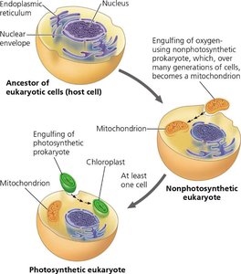
Q7. Describe the endosymbiosis theory and four pieces of evidence supporting it.
Background
Topic: Evolution and Endosymbiosis Theory
This question tests your understanding of how eukaryotic cells may have evolved from prokaryotic ancestors.
Key Terms:
Endosymbiosis Theory: Eukaryotic cells originated from prokaryotic cells engulfing other prokaryotes.
Mitochondria and Chloroplasts: Organelles believed to have originated from free-living bacteria.
Step-by-Step Guidance
Review the diagram showing the process of endosymbiosis.
List features mitochondria and chloroplasts share with prokaryotes (e.g., DNA, double membranes).
Identify four pieces of evidence supporting the theory (e.g., own DNA, ribosomes, reproduction, similarity to bacteria).
Explain how these features support the theory.
Try solving on your own before revealing the answer!
Final Answer:
The endosymbiosis theory proposes that mitochondria and chloroplasts originated from prokaryotic cells engulfed by ancestral eukaryotes. Evidence includes: (1) both have their own DNA, (2) both have double membranes, (3) both reproduce independently, (4) both have ribosomes similar to prokaryotes.
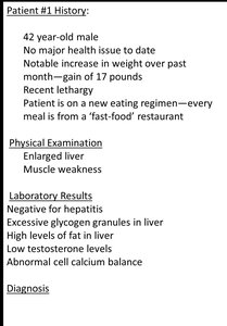
Q8. Predict the malfunctioning organelle in a patient with excessive glycogen granules, high fat in liver, and abnormal cell calcium balance.
Background
Topic: Organelle Function and Disease
This question tests your ability to connect symptoms to organelle malfunction.
Key Terms:
Glycogen Granules: Storage form of glucose in liver.
Fat Accumulation: Indicates issues with lipid metabolism.
Calcium Balance: Related to smooth ER function.
Step-by-Step Guidance
Analyze the symptoms: enlarged liver, muscle weakness, high fat, abnormal calcium.
Recall which organelle is involved in lipid metabolism and calcium storage (smooth ER).
Consider how malfunction of this organelle could lead to these symptoms.
Review the role of the smooth ER in detoxification, lipid synthesis, and calcium storage.
Try solving on your own before revealing the answer!
Final Answer:
The malfunctioning organelle is the smooth endoplasmic reticulum (SER), which is responsible for lipid metabolism, detoxification, and calcium storage. Dysfunction in the SER can lead to fat accumulation, abnormal calcium balance, and excessive glycogen storage.
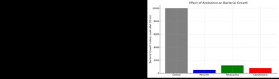
Q9. What are the independent and dependent variables in the experiment testing antibiotic effectiveness on bacterial growth?
Background
Topic: Experimental Design
This question tests your ability to identify variables in a scientific experiment.
Key Terms:
Independent Variable: The factor manipulated by the experimenter (type of antibiotic).
Dependent Variable: The factor measured (bacterial growth).
Step-by-Step Guidance
Review the experiment setup: different antibiotics are tested.
Identify what is being changed (independent variable).
Identify what is being measured (dependent variable).
Check the bar graph for clarity on these variables.
Try solving on your own before revealing the answer!
Final Answer:
The independent variable is the type of antibiotic used. The dependent variable is the amount of bacterial growth measured after treatment.
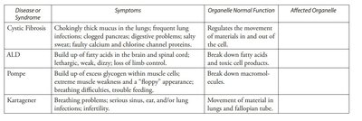
Q10. What organelles are likely malfunctioning in diseases such as cystic fibrosis, ALD, Pompe, and Kartagener syndrome?
Background
Topic: Organelle Dysfunction and Disease
This question tests your ability to match symptoms to affected organelles.
Key Terms:
Cystic Fibrosis: Issues with plasma membrane and channel proteins.
ALD: Issues with peroxisomes (fatty acid breakdown).
Pompe: Issues with lysosomes (macromolecule breakdown).
Kartagener: Issues with cilia (microtubules).
Step-by-Step Guidance
Read the symptoms and normal function for each disease.
Recall which organelle is responsible for each function.
Match the symptoms to the malfunctioning organelle.
Review the table for confirmation.
Try solving on your own before revealing the answer!
Final Answer:
Cystic Fibrosis: Plasma membrane; ALD: Peroxisomes; Pompe: Lysosomes; Kartagener: Cilia (microtubules). Each disease is linked to dysfunction in the organelle responsible for the affected process.
