 Back
Cell Structure, Metabolism, Cellular Respiration, and Photosynthesis: Exam #2 Study Guide
Study Guide - Smart Notes
 Tailored notes based on your materials, expanded with key definitions, examples, and context.
Chapter 6: A Tour of the Cell
Microscopy
Microscopy is essential for visualizing cells and their internal structures. Different types of microscopes provide varying levels of resolution and magnification.
Micrometer (μm): Unit of measurement for cell structures (1 μm = 10-6 meters).
Light Microscope: Uses visible light to observe cells; can resolve structures down to about 200 nm.
Electron Microscopy: Uses electron beams for much higher resolution (up to 0.2 nm); reveals ultrastructure of cells.
Resolution: Ability to distinguish two points as separate.
Magnification: Enlargement of the image.
What can be seen: Light microscopes show cell walls, nuclei, and some organelles; electron microscopes reveal ribosomes, membranes, and detailed organelle structure.
Prokaryotic Cell Structure
Prokaryotic cells lack membrane-bound organelles and are structurally simpler than eukaryotes.
Cytoplasm: The region inside the plasma membrane containing cytosol and cellular components.
Nucleoid: Area where the DNA is located, not membrane-bound.
Ribosome: Site of protein synthesis.
Plasma Membrane: Selectively permeable barrier.
Cell Wall: Provides structural support.
Flagella: Used for movement.
Eukaryotic Cell Structure
Eukaryotic cells contain membrane-bound organelles and are generally larger than prokaryotic cells.
Organelles: Specialized structures (e.g., nucleus, mitochondria, ER).
Surface Area to Volume Ratio: Limits cell size; eukaryotes evolved internal membranes to increase surface area.
Nucleus
The nucleus is the control center of the cell, housing genetic material and coordinating cellular activities.
Genome: Complete set of genes.
Chromatin: DNA and protein complex; condenses to form chromosomes.
Nuclear Envelope: Double membrane with nuclear pores for transport.
Nucleolus: Site of ribosome synthesis.
Ribosomes
Ribosomes are responsible for protein synthesis and can be free in the cytosol or bound to the ER.
rRNA: Ribosomal RNA, structural and catalytic component.
Large and Small Subunits: Two parts of the ribosome.
Free vs. Bound Ribosomes: Free ribosomes synthesize cytosolic proteins; bound ribosomes synthesize proteins for membranes or secretion.
Endomembrane System
The endomembrane system coordinates the synthesis, modification, and transport of proteins and lipids.
Components: Includes the ER (smooth and rough), Golgi apparatus, lysosomes, vacuoles, and transport vesicles.
Smooth ER: Lipid synthesis, detoxification.
Rough ER: Protein synthesis (with ribosomes).
Golgi Apparatus: Modifies, sorts, and packages proteins.
Transport Vesicles: Move materials between organelles.
Lysosome: Contains hydrolytic enzymes for digestion.
Vacuoles: Storage and structural support (central vacuole in plants).
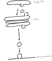
Secretory Pathway Diagram
The secretory pathway describes the movement of proteins from their synthesis in the rough ER, through the Golgi apparatus, and finally to the plasma membrane for secretion or membrane insertion.
Soluble glycoproteins: Synthesized in the rough ER, modified in the Golgi, and secreted out of the cell.
Transmembrane glycoproteins: Inserted into the plasma membrane, with carbohydrates facing the extracellular side.
Mitochondria
Mitochondria are the site of cellular respiration, converting chemical energy in food to ATP.
Structure: Double membrane, matrix, cristae.
Endosymbiont Theory: Originated from engulfed prokaryotes.
Chloroplast
Chloroplasts are the site of photosynthesis in plants and algae.
Structure: Double membrane, thylakoid membranes, stroma.
Endosymbiont Theory: Originated from engulfed photosynthetic prokaryotes.
Peroxisome
Peroxisomes contain enzymes that break down hydrogen peroxide and other toxic substances.
Hydrogen Peroxide: Byproduct of metabolic reactions, broken down by catalase.
Cytoskeleton
The cytoskeleton provides structural support, facilitates movement, and organizes cell contents.
Microtubules: Tubulin polymers, involved in cell shape, transport, and division.
Microfilaments (Actin): Actin polymers, involved in movement and contraction.
Intermediate Filaments: Provide mechanical strength.
Extracellular Matrix (ECM) and Cell Junctions
The ECM and cell junctions maintain tissue structure and facilitate communication between cells.
ECM: Network of proteins and carbohydrates outside animal cells.
Cell Junctions: Tight junctions, desmosomes, gap junctions, plasmodesmata.
Chapter 8: Introduction to Metabolism
Organization of Metabolism
Metabolism encompasses all chemical reactions in the cell, organized into metabolic pathways.
Catabolic Pathways: Break down molecules, release energy (downhill reactions).
Anabolic Pathways: Build molecules, require energy (uphill reactions).
Energy and Thermodynamics
Energy is the capacity to do work, and cells obey the laws of thermodynamics.
First Law: Energy cannot be created or destroyed.
Second Law: Entropy increases in spontaneous processes.
Types of Energy: Potential, kinetic, chemical, light, thermal.
Chemical Reactions in the Cell
Cellular reactions involve changes in free energy and are catalyzed by enzymes.
Free Energy (G): Energy available to do work.
Change in Free Energy (ΔG): Determines spontaneity.
Exergonic Reactions: Release energy (ΔG < 0).
Endergonic Reactions: Require energy (ΔG > 0).
ATP: Main energy currency.
Activation Energy (EA): Energy barrier to reaction.
Enzymes: Biological catalysts, lower EA.
Factors Influencing Enzyme Activity
Enzyme activity is affected by temperature, pH, cofactors, and inhibitors.
Competitive Inhibition: Inhibitor binds active site.
Noncompetitive Inhibition: Inhibitor binds allosteric site.
Chapter 9: Cellular Respiration and Fermentation
Oxidation-Reduction Reactions (Redox)
Cellular respiration involves redox reactions, transferring electrons and energy.
Oxidation: Loss of electrons.
Reduction: Gain of electrons.
Electron Carriers: NAD+/NADH, FAD/FADH2.
Glycolysis
Glycolysis is the first stage of cellular respiration, occurring in the cytosol.
Energy Investment Phase: Uses ATP to split glucose.
Energy Payoff Phase: Produces ATP and NADH.
Net Output: 2 ATP, 2 NADH, 2 pyruvate per glucose.
Pyruvate Oxidation
Pyruvate is converted to Acetyl CoA in the mitochondria, producing NADH and CO2.
Equation:
Citric Acid Cycle
The citric acid cycle completes the oxidation of glucose, generating NADH, FADH2, ATP, and CO2.
Location: Mitochondrial matrix.
Purpose: Energy extraction and electron carrier production.
Oxidative Phosphorylation
Oxidative phosphorylation uses the electron transport chain and chemiosmosis to produce ATP.
Electron Transport Chain: Series of complexes in the inner mitochondrial membrane.
Proton Gradient: Drives ATP synthesis via ATP synthase.
ATP Yield: About 34 ATP per glucose.
Anaerobic Respiration and Fermentation
Fermentation allows ATP production without oxygen, regenerating NAD+.
Alcohol Fermentation: Produces ethanol and CO2.
Lactic Acid Fermentation: Produces lactic acid.
ATP Yield: Much lower than aerobic respiration.
Chapter 10: Photosynthesis
How Organisms Obtain Energy
Organisms are classified by how they obtain energy: heterotrophs consume organic molecules, autotrophs produce their own.
Photoautotrophs: Use light energy to synthesize organic molecules.
Overview of Photosynthesis
Photosynthesis converts solar energy into chemical energy, producing glucose and oxygen.
Overall Reaction:
Oxidized: Water.
Reduced: Carbon dioxide.
Endergonic Reaction: Requires energy input.
Chloroplast Structure
Chloroplasts contain thylakoid membranes, grana, and stroma, where photosynthesis occurs.
Chlorophyll: Located in thylakoid membranes.
Photosystems: Embedded in thylakoid membranes.
Calvin Cycle: Occurs in the stroma.
Light Reactions
Light reactions capture solar energy to produce ATP and NADPH.
Photosystem II: Absorbs light, splits water.
Electron Transport Chain: Transfers electrons, pumps protons.
Photophosphorylation: ATP synthesis via ATP synthase.
Photosystem I: Produces NADPH.
The Calvin Cycle
The Calvin cycle uses ATP and NADPH to fix carbon dioxide and produce glucose.
Carbon Fixation: CO2 attached to RuBP by Rubisco.
Reduction: 3-PGA converted to G3P.
Regeneration: RuBP is regenerated.
Comparison: Oxidative Phosphorylation vs. Photophosphorylation
Both processes use electron transport chains and ATP synthase, but differ in energy source and location.
Oxidative Phosphorylation: Occurs in mitochondria, uses chemical energy from food.
Photophosphorylation: Occurs in chloroplasts, uses light energy.
Additional info:
Some explanations and diagrams were expanded for clarity and completeness.
