 Back
Cell Structures: Size, Visualization, and Prokaryotic Structures
Study Guide - Smart Notes
 Tailored notes based on your materials, expanded with key definitions, examples, and context.
Cell Structures: Size, Visualization, and Prokaryotic Structures
Why Are Cells So Small?
Cells are the fundamental units of life, and their small size is crucial for efficient biological function. The surface area-to-volume ratio is a key factor that limits cell size, as it affects the rate at which materials can enter or exit the cell.
Surface Area-to-Volume Ratio: As a cell increases in size, its volume grows faster than its surface area. This ratio determines how efficiently a cell can exchange nutrients and waste with its environment.
Diffusion Efficiency: Smaller cells have a higher surface area relative to their volume, allowing for more efficient diffusion of substances.
Multicellularity: Large organisms, such as elephants, are composed of many small cells rather than a single large cell to maintain efficient metabolic exchange.
Example: If you compare dissolving large and small candies, the smaller candies dissolve faster due to their greater surface area relative to volume. Similarly, smaller cells can more rapidly exchange materials.


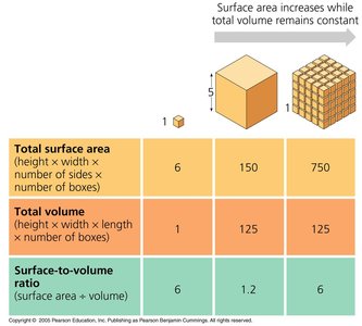
Additional info: The mathematical relationship for surface area-to-volume ratio is given by:
Cell Size and Visualization
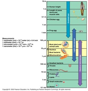
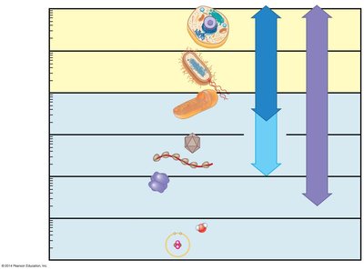
Cells and their components vary greatly in size, from atoms and small molecules to entire cells. Different types of microscopes are used to visualize these structures, depending on their scale.
Scale of Biological Structures: Most plant and animal cells are between 10 and 100 micrometers (μm) in diameter. Organelles, viruses, and molecules are much smaller.
Microscopy: Light microscopes are used for cells and larger organelles, while electron microscopes are required for smaller structures such as viruses and ribosomes.
Measurement Units: Common units include centimeters (cm), millimeters (mm), micrometers (μm), and nanometers (nm).
Microscopy Techniques
Microscopes are essential tools for studying cell structure. Different types of microscopy provide varying levels of detail and contrast.
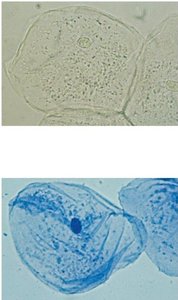
Light Microscopy: Uses visible light to observe cells and tissues. Staining can enhance contrast and reveal specific structures.
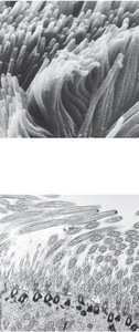
Scanning Electron Microscopy (SEM): Provides detailed surface images of cells and organelles by scanning with a focused beam of electrons.
Transmission Electron Microscopy (TEM): Reveals internal structures by transmitting electrons through thin sections of specimens.
Super-Resolution Microscopy: Advanced techniques that surpass the resolution limits of conventional light microscopy.
Prokaryotic and Eukaryotic Cells: Common Features
All cells share certain fundamental features, regardless of their classification as prokaryotic or eukaryotic.
Plasma Membrane: A selective barrier that encloses the cell.
Cytosol: The semifluid substance within the cell where metabolic reactions occur.
Chromosomes: Structures containing genetic material (DNA).
Ribosomes: Complexes that synthesize proteins.
Prokaryotic Cell Structure
Prokaryotic cells, such as bacteria, have distinct structural features that differentiate them from eukaryotic cells.
Size: Prokaryotic cells are generally smaller than eukaryotic cells.
Membrane-Bound Organelles: Prokaryotes lack membrane-bound organelles such as nuclei and mitochondria.
DNA Location: DNA is located in the nucleoid region, not enclosed by a membrane.
Other Features: Prokaryotes may have a cell wall, capsule, fimbriae, and flagella for protection, attachment, and movement.
Example: The bacterium Corynebacterium diphtheriae shows typical prokaryotic features such as a nucleoid, ribosomes, and cell wall.
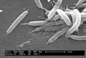
Additional info: Prokaryotic cells are classified into two domains: Bacteria and Archaea.
Visualizing Eukaryotic Cells
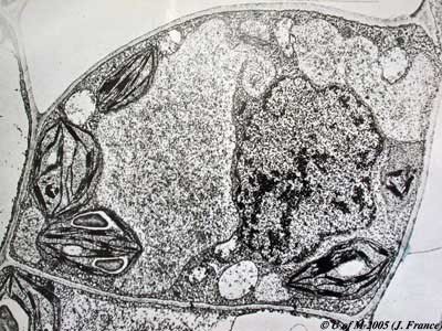
Eukaryotic cells contain membrane-bound organelles, including a nucleus, mitochondria, and chloroplasts (in plants). These structures are visible under advanced microscopy techniques.
Nucleus: Contains the cell's genetic material and is surrounded by a nuclear envelope.
Mitochondria: Organelles responsible for energy production.
Chloroplasts: Organelles in plant cells responsible for photosynthesis.
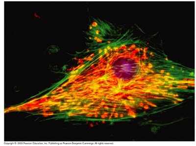
Cytoskeleton: Network of fibers that provides structural support and facilitates movement.
 ----------------------------------------
