 Back
Cell–Cell Interactions: Structure, Adhesion, and Communication
Study Guide - Smart Notes
 Tailored notes based on your materials, expanded with key definitions, examples, and context.
Cell–Cell Interactions
Introduction
Cell–cell interactions are fundamental for the structure and function of multicellular organisms. These interactions involve the extracellular matrix, cell adhesion structures, and communication pathways that allow cells to coordinate their activities. This chapter explores the molecular and structural basis of cell–cell interactions in both animal and plant cells.
The Extracellular Matrix (ECM) of Animal Cells
Structure and Function of the ECM
The extracellular matrix (ECM) is a complex network of proteins and polysaccharides secreted by animal cells. It provides structural support, segregates tissues, and regulates intercellular communication.
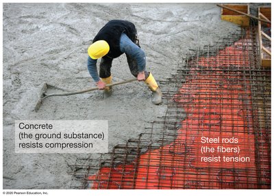
Fibrous component: Mainly composed of collagen, which forms strong, rope-like fibers that resist tension (pulling forces).
Ground substance: Made of proteoglycans—proteins attached to many polysaccharides—giving the ECM a gel-like, compressive resistance.
Collagen molecules twist into triple helices, which then group together to form fibrils and larger fibers.
Proteoglycans are responsible for the rubber-like consistency of cartilage and other connective tissues.
Example: The ECM in cartilage provides both tensile strength (from collagen) and compressive resistance (from proteoglycans), similar to how steel rods and concrete work together in reinforced concrete.

Indirect Cell–Cell Attachments in Plants
The Middle Lamella
Plant cells are held together by a layer called the middle lamella, which is rich in pectins (gelatinous polysaccharides). This layer glues adjacent plant cells together and is continuous with their cell walls.
Middle lamella: Acts as a glue, holding plant cells together and allowing the formation of tissues.
Pectins: The main component, providing a gel-like consistency that helps resist compression and maintain tissue integrity.

Cell–Cell Adhesion and Communication in Animal Cells
Overview of Cell Junctions
Animal cells use specialized structures for adhesion and communication. These include tight junctions, desmosomes, and gap junctions.
Tight junctions: Seal cells together, preventing leakage of extracellular fluid.
Desmosomes: Connect the cytoskeletons of adjacent cells, providing strong adhesion.
Gap junctions: Act as channels for communication between cells, allowing ions and small molecules to pass directly from one cell to another.

Tight Junctions
Tight junctions are composed of membrane proteins in adjacent animal cells that line up and bind to each other, forming a watertight seal. They are commonly found in epithelial tissues and can regulate the passage of substances between cells.
Prevent leakage of extracellular fluid between cells.
May loosen to permit selective transport of molecules.

Desmosomes
Desmosomes are strong cell–cell attachments found in animal epithelial and muscle cells. They consist of linking proteins and cytosolic anchoring proteins, which are reinforced by intermediate filaments of the cytoskeleton.
Provide mechanical strength to tissues by connecting the cytoskeletons of adjacent cells.
Intermediate filaments attach to anchoring proteins inside the cell, stabilizing the structure.

Gap Junctions
Gap junctions are protein channels that connect adjacent animal cells, allowing the flow of ions and small molecules. These communication portals help coordinate cellular activities and enable rapid passage of regulatory signals.
Allow direct communication between cells in the same tissue.
Can regulate gene expression and protein activity by transmitting signals.

Cell–Cell Communication in Plants
Plasmodesmata
Plant cells are connected by plasmodesmata, which are membrane-lined channels that traverse the cell wall. These channels allow the plasma membranes, cytoplasm, and smooth endoplasmic reticulum (ER) of adjacent cells to connect.
Symplast: The shared cytoplasm connected by plasmodesmata.
Apoplast: The extracellular space outside the plasma membrane.


Cell–Cell Signaling in Multicellular Organisms
Signaling Molecules and Their Actions
Cells communicate over short and long distances using signaling molecules such as neurotransmitters and hormones.
Neurotransmitters: Open or close ion channels in target cells, enabling rapid communication over short distances.
Hormones: Information-carrying molecules secreted into the body, acting on distant target cells to coordinate physiological responses.

Signal Reception and Transduction
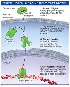
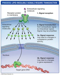
Cell signaling involves the reception of a signal by a receptor, followed by transduction and response. Signals can be lipid-soluble (e.g., steroid hormones) or lipid-insoluble (e.g., peptide hormones).
Lipid-soluble signals: Pass through the plasma membrane and bind to intracellular receptors, often affecting gene expression directly.
Lipid-insoluble signals: Bind to cell surface receptors, triggering a cascade of intracellular events (signal transduction) that may amplify and diversify the signal.
Signal Amplification and Diversification
Signal transduction pathways can amplify and diversify the original signal. Two major systems are:
G-protein-coupled receptors (GPCRs): Initiate the production of intracellular second messengers, amplifying the signal and leading to multiple cellular responses.
Enzyme-linked receptors: Phosphorylate proteins inside the target cell, often triggering a phosphorylation cascade that amplifies the signal.


Crosstalk Between Signaling Pathways
Different signaling pathways can interact with each other, a phenomenon known as crosstalk. This allows cells to integrate multiple signals and coordinate complex responses.
Additional info: The ECM, cell junctions, and signaling pathways are essential for tissue structure, integrity, and coordinated function in multicellular organisms. Disruption of these systems can lead to diseases such as cancer, where cell adhesion and communication are often altered.
