 Back
BackCell–Cell Interactions: Structure, Adhesion, and Communication
Study Guide - Smart Notes
 Tailored notes based on your materials, expanded with key definitions, examples, and context.
Cell–Cell Interactions
Introduction
Cell–cell interactions are fundamental for the structure and function of multicellular organisms. These interactions involve the extracellular matrix, specialized cell junctions, and signaling mechanisms that allow cells to adhere, communicate, and coordinate activities.
The Extracellular Matrix (ECM) of Animal Cells
Structure and Function
Extracellular Matrix (ECM): A fiber composite secreted by most animal cells, providing structural support and mediating cell signaling.
Fibrous Component: Primarily composed of collagen, which forms triple helices that aggregate into strong collagen fibrils.
Ground Substance: Made of proteoglycans—proteins attached to many polysaccharides—giving tissues like cartilage a rubber-like consistency.
Example: The ECM is crucial in connective tissues such as tendons and cartilage, where it resists tension and compression.
Indirect Cell–Cell Attachments in Plants
The Middle Lamella
Middle Lamella: A central layer of gelatinous pectins that glues adjacent plant cells together and is continuous with their cell walls.
Provides mechanical stability and enables the formation of plant tissues.
Example: The middle lamella is essential for the integrity of plant tissues, especially during growth and development.
Cell–Cell Adhesion and Communication in Animal Cells
Tight Junctions
Tight junctions are specialized cell–cell attachments that create a watertight seal between adjacent animal cells, particularly in epithelial tissues.
Structure: Composed of membrane proteins that line up and bind to each other, stitching cells together.
Function: Prevents leakage of extracellular fluid and may loosen to permit selective transport.
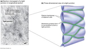
Example: Tight junctions are found in the lining of the intestines, preventing the passage of harmful substances between cells.
Desmosomes
Desmosomes: Strong cell–cell attachments common in epithelial and muscle cells.
Composed of linking proteins and cytosolic anchoring proteins, reinforced by cytoskeletal intermediate filaments.
Provide mechanical strength by binding the cytoskeletons of adjacent cells together.
Example: Desmosomes are abundant in tissues subject to mechanical stress, such as the skin and heart muscle.
Gap Junctions
Gap Junctions: Protein channels that connect adjacent animal cells, allowing the flow of ions and small molecules.
Enable rapid communication and coordination of cellular activities, such as in cardiac muscle contraction.
Example: Gap junctions synchronize heart muscle contractions by allowing ions to pass quickly between cells.
Cell–Cell Communication in Plants
Plasmodesmata
Plasmodesmata: Membrane-lined channels that traverse plant cell walls, connecting the plasma membranes, cytoplasm, and smooth endoplasmic reticulum (ER) of adjacent cells.
Divide plant tissues into two compartments:
Symplast: Shared cytoplasm connected by plasmodesmata.
Apoplast: Extracellular space outside the plasma membrane.
Example: Plasmodesmata facilitate the movement of nutrients and signaling molecules between plant cells.
Cell–Cell Signaling in Multicellular Organisms
Signaling Molecules and Mechanisms
Neurotransmitters: Chemical messengers that open or close ion channels in distant cells, enabling rapid communication in nervous tissue.
Hormones: Information-carrying molecules secreted by cells, circulating throughout the body to act on distant target cells.
Example: Insulin is a hormone that regulates glucose uptake in cells throughout the body.
Signal Transduction Pathways
Signal transduction converts an extracellular signal into an intracellular response, often amplifying and diversifying the message.
Two major types of signal transduction systems:
G-Protein-Coupled Receptors (GPCRs): Initiate the production of intracellular second messengers, amplifying and diversifying the signal.
Enzyme-Linked Receptors: Trigger phosphorylation cascades by phosphorylating proteins inside the target cell.
Example: The adrenaline signaling pathway uses GPCRs to rapidly mobilize energy stores in response to stress.
Integration of Signaling Pathways
Signaling pathways often interact through crosstalk, allowing cells to integrate multiple signals and coordinate complex responses.
Example: Crosstalk between growth factor and stress response pathways can determine whether a cell divides or initiates repair mechanisms.
