 Back
BackCellular Membranes, Diffusion, and Organelles: Structure and Function
Study Guide - Smart Notes
 Tailored notes based on your materials, expanded with key definitions, examples, and context.
Cellular Membranes: Structure and Function
Overview of Cell Membranes
Cell membranes are fundamental to all living organisms, providing boundaries, structural integrity, and selective permeability. The plasma membrane surrounds every cell, separating the internal environment from the external surroundings and regulating the movement of substances in and out.
Plasma Membrane: A dynamic, complex structure composed primarily of lipids and proteins.
Functions: Maintains cell shape, mediates communication, and controls transport.
Membrane Fluidity: Essential for proper function, influenced by lipid composition and cholesterol.
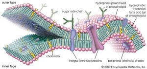
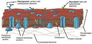
Phospholipid Bilayer
The plasma membrane is primarily composed of a phospholipid bilayer, which forms the basic structural framework. Phospholipids have both hydrophilic (water-loving) heads and hydrophobic (water-fearing) tails, resulting in a bilayer arrangement.
Hydrophilic Head: Faces outward toward aqueous environments.
Hydrophobic Tails: Face inward, away from water.
Bilayer Arrangement: Creates a semi-permeable barrier.

Components of the Cell Membrane
In addition to phospholipids, cell membranes contain proteins, cholesterol, glycolipids, and glycoproteins. These components contribute to membrane structure, fluidity, and function.
Proteins: Integral and peripheral proteins perform transport, signaling, and structural roles.
Cholesterol: Modulates membrane fluidity and stability.
Glycolipids and Glycoproteins: Involved in cell recognition and signaling.
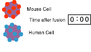
Fluid Mosaic Model
The fluid mosaic model describes the cell membrane as a dynamic structure where lipids and proteins move laterally within the bilayer. This fluidity is crucial for membrane function, including transport and cell signaling.
Experiment: Fusion of human and mouse cells with labeled surface proteins demonstrated lateral diffusion of membrane components.
Membrane Fluidity: Influenced by lipid saturation and cholesterol content.
Lipid Composition and Membrane Fluidity
Membrane fluidity depends on the types of lipids present. Saturated fatty acids pack tightly, reducing fluidity, while unsaturated fatty acids introduce kinks, increasing fluidity. Cholesterol acts as a buffer, stabilizing membrane fluidity under varying conditions.
Saturated Lipids: No double bonds, straight chains, less fluid.
Unsaturated Lipids: One or more double bonds, kinked chains, more fluid.
Cholesterol: Inserts between phospholipids, modulating fluidity.
Transport Across Cell Membranes
Diffusion and Osmosis
Diffusion is the passive movement of molecules from areas of high concentration to low concentration. Osmosis is the passive movement of water across membranes toward areas of higher solute concentration.
Simple Diffusion: Movement of membrane-permeable substances without energy input.
Facilitated Diffusion: Movement of low or non-permeable substances via transport proteins.
Osmosis: Water moves toward higher solute concentration.
Types of Solutions
Cells can be exposed to hypotonic, isotonic, or hypertonic solutions, affecting water movement and cell volume.
Hypotonic: Lower solute concentration outside the cell; water enters the cell.
Isotonic: Equal solute concentration; no net water movement.
Hypertonic: Higher solute concentration outside; water leaves the cell.
Transport Proteins
Non-permeable substances cross the membrane via transport proteins, which can be channels or carriers. Transport can be passive (no energy required) or active (requires energy).
Channel Proteins: Form pores for specific molecules.
Carrier Proteins: Bind and transport substances across the membrane.
Active Transport: Moves substances against concentration gradients using ATP.
Cellular Organelles and Internal Membranes
Eukaryotic vs. Prokaryotic Cells
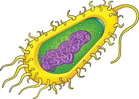
Cells are classified as prokaryotic or eukaryotic based on internal structure. Eukaryotic cells have membrane-bound organelles, while prokaryotic cells lack them.
Prokaryotes: No internal membranes, unicellular, single circular chromosome.
Eukaryotes: Internal membranes, uni- or multicellular, multiple linear chromosomes, specialized structures.
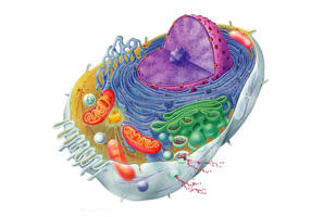
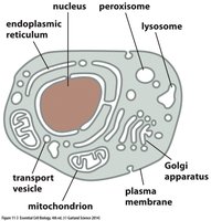
Major Eukaryotic Organelles
Eukaryotic cells contain several membrane-bound organelles, each with specialized functions.
Nucleus: Stores and protects chromosomes; site of transcription.
Endoplasmic Reticulum (ER): Synthesizes and packages proteins and lipids.
Golgi Apparatus: Modifies, sorts, and packages proteins for export.
Mitochondria: Produces ATP via cellular respiration.
Chloroplasts: Converts sunlight to chemical energy (in plants).
Lysosomes: Breaks down macromolecules with enzymes.
Endosymbiosis Theory
The theory of endosymbiosis explains the origin of mitochondria and chloroplasts in eukaryotic cells, proposing that these organelles originated from symbiotic prokaryotes.
Mitochondria and Chloroplasts: Contain their own DNA, double membranes, and reproduce independently.
Cytoskeleton and Intracellular Transport
Cytoskeleton Structure and Function
The cytoskeleton is a network of protein filaments that provides structural support, facilitates cell movement, and enables intracellular transport.
Microtubules: Largest filaments, radiate from a central point, drive mitosis and meiosis.
Intermediate Filaments: Rope-like, provide mechanical strength.
Microfilaments: Smallest, important for cell shape and movement.
Motor Proteins and Vesicle Transport
Motor proteins move organelles and vesicles along cytoskeletal tracks, facilitating intracellular transport and communication.
Motor Proteins: Use ATP to transport cargo along microtubules and microfilaments.
Membrane Transport: Endocytosis and Exocytosis
Endocytosis and Exocytosis
Cells use membranes to engulf (endocytosis) or release (exocytosis) large particles and molecules. These processes are organized by the cytoskeleton and are essential for nutrient uptake, waste removal, and signaling.
Endocytosis: Membrane surrounds and encloses substances for internalization.
Exocytosis: Membranes fuse to eject contents outside the cell.
Lysosomes: Break down engulfed particles with enzymes.
Receptor-Mediated Endocytosis: Selectively internalizes specific molecules.
Summary Table: Comparison of Prokaryotic and Eukaryotic Cells
Feature | Prokaryotes | Eukaryotes |
|---|---|---|
Internal Membranes | Absent | Present |
Nucleus | Absent | Present |
Cell Wall | Complex | Simpler or absent |
Chromosomes | Single, circular | Multiple, linear |
Organisms | Bacteria, Archaea | Animals, Plants, Fungi, Protists |
Key Equations
Diffusion: Fick's Law of Diffusion Where J is the flux, D is the diffusion coefficient, and \frac{dC}{dx} is the concentration gradient.
Osmosis: Osmotic Pressure Where \Pi is osmotic pressure, i is the van 't Hoff factor, M is molarity, R is the gas constant, and T is temperature.
Additional info: Academic context was added to clarify the structure and function of membranes, organelles, and transport mechanisms, as well as to provide definitions and examples for key terms.
