 Back
BackCellular Processes: Protein Activity, Nuclear Transport, Endomembrane System, and Cytoskeleton
Study Guide - Smart Notes
 Tailored notes based on your materials, expanded with key definitions, examples, and context.
Cell Processes and Lab Techniques
Differential Centrifugation
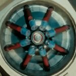
Differential centrifugation is a laboratory technique used to separate cellular components based on their density. This method is fundamental for isolating organelles and studying their functions in cell biology.
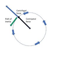
Principle: Centrifugal force separates particles by density; heavier components form a pellet, lighter remain in the supernatant.
Applications: Used to isolate nuclei, mitochondria, lysosomes, and other organelles for biochemical analysis.
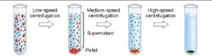
Process: Sequential spins at increasing speeds allow stepwise separation of cellular fractions.
Example: Low-speed centrifugation pellets nuclei; higher speeds pellet mitochondria, lysosomes, etc.

Protein Structure and Function
Protein Binding and Shape Change
Proteins are highly specific biological machines whose activity depends on their ability to bind substrates and undergo conformational changes. This specificity is crucial for cellular processes such as signaling, transport, and catalysis.
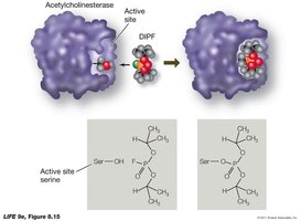
Binding Specificity: Proteins bind ligands, substrates, or signals via complementary chemical interactions (e.g., enzyme-substrate, receptor-signal).
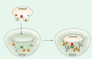
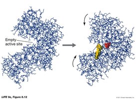
Shape Change: Binding often induces a conformational change, altering protein activity.
Key Terms: Active site (region of enzyme where substrate binds), ligand (molecule that binds to a protein).
Example: Enzyme active site changes shape upon substrate binding, facilitating catalysis.
Nuclear Transport
Nuclear Pore Complex and Signals
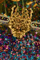
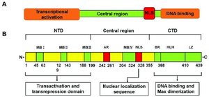
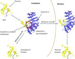
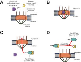
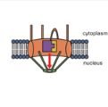
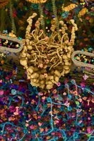
The nuclear pore complex (NPC) regulates the entry and exit of molecules between the cytoplasm and nucleus. Proteins require specific signals to be transported across the nuclear envelope.
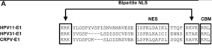
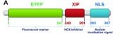
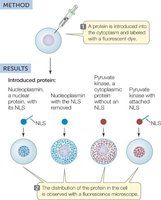
Nuclear Localizing Signal (NLS): A sequence of amino acids that directs proteins to the nucleus.
Nuclear Exit Signal (NES): A different sequence that facilitates export from the nucleus.
Shuttle Proteins: Bind to NLS and transport cargo through the NPC.
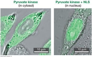
Example: Adding an NLS to a cytoplasmic protein causes it to localize in the nucleus.


Endomembrane System and Protein Sorting
Protein Synthesis and Sorting
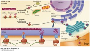
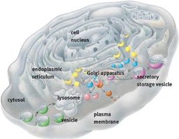
Proteins are synthesized on ribosomes and sorted to their correct cellular locations by signal sequences. The endomembrane system includes the ER, Golgi apparatus, lysosomes, and vesicles.
Cytosolic Ribosomes: Synthesize proteins for cytosol, mitochondria, chloroplasts, peroxisomes, and nucleus.
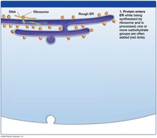
RER Ribosomes: Synthesize proteins for secretion, membrane, lysosomes, and ER.
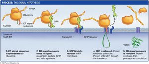
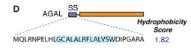
ER Signal Sequence: Directs ribosome to the rough ER for co-translational import.
Example: Proteins with ER signal sequence are fed into the ER membrane during synthesis.
Pathway from ER to Outside of Cell
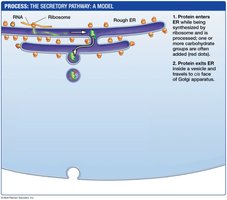
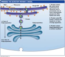
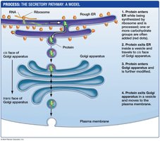
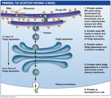
Proteins destined for secretion or membrane insertion follow a pathway from the ER to the Golgi apparatus, then to the plasma membrane or lysosome via vesicular transport.
Vesicular Transport: Proteins are packaged into vesicles that bud from the ER and fuse with the Golgi.
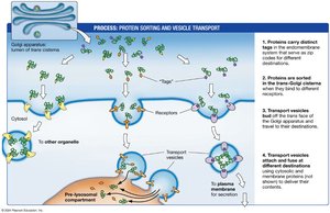
Sorting in Golgi: Proteins are modified and sorted based on amino acid tags.
Secretion: Vesicles fuse with the plasma membrane, releasing proteins outside the cell.
Protein Sorting and Vesicle Formation
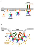
Specific amino acid tags direct proteins to their final destinations. Integral membrane proteins in the Golgi recognize these tags and facilitate vesicle formation.
Tag Recognition: Golgi membrane proteins bind to sorting sequences.
Vesicle Formation: Clusters of tagged proteins are packaged into vesicles by "pinching off" the membrane.
Membrane Transport: Vesicles and Lysosomes
Vesicular Transport Mechanisms
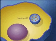
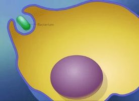
Cells use vesicles to transport molecules across membranes. This includes endocytosis, exocytosis, pinocytosis, and phagocytosis.
Pinocytosis: Uptake of small dissolved substances or fluids.
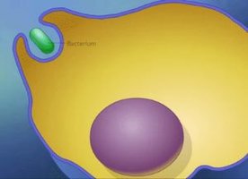
Phagocytosis: Engulfment of large particles or entire cells.
Exocytosis: Release of molecules to extracellular space.
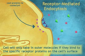
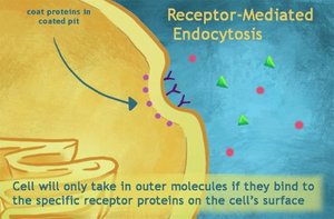
Endocytosis: Receptor-mediated uptake of specific molecules.

Pathways to the Lysosome
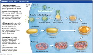
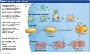
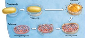
Lysosomes are cellular organelles responsible for degrading and recycling materials. There are three main pathways to the lysosome: receptor-mediated endocytosis, phagocytosis, and autophagy.
Receptor-Mediated Endocytosis: Molecules bind to cell surface receptors and are internalized.
Phagocytosis: Entire cells or large particles are engulfed and degraded.
Autophagy: Worn-out organelles are enclosed in vesicles and digested.
Cytoskeleton: Structure and Function
Cytoskeletal Filaments
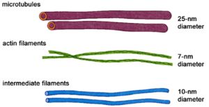
The cytoskeleton is a network of protein filaments that provides structural support, facilitates movement, and organizes cellular components. Most cells possess a cytoskeleton composed of actin filaments, intermediate filaments, and microtubules.
Actin Filaments: Thin, flexible fibers involved in cell shape, movement, and division.
Intermediate Filaments: Provide mechanical strength and anchor organelles.
Microtubules: Hollow tubes that support cell shape, transport vesicles, and form cilia/flagella.
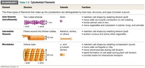
Filament | Subunits | Functions |
|---|---|---|
Actin Filaments | Actin | Cell shape, movement, division |
Intermediate Filaments | Keratin, lamins | Mechanical strength, organelle anchoring |
Microtubules | α- and β-tubulin | Cell shape, vesicle transport, cilia/flagella |
Motor Proteins and Cellular Movement
Motor proteins such as myosin, kinesin, and dynein interact with cytoskeletal filaments to generate movement within cells.
Myosin: Pulls on actin filaments for muscle contraction and cell movement.
Kinesin and Dynein: Move along microtubules, transporting vesicles and organelles.
Cilia and Flagella: Motor proteins drive movement of these structures for cell motility.

Summary Table: Cytoskeletal Filaments
Type | Diameter | Subunit | Function |
|---|---|---|---|
Actin Filaments | 7 nm | Actin | Cell shape, movement, division |
Intermediate Filaments | 10 nm | Keratin, lamins | Mechanical strength, anchoring |
Microtubules | 25 nm | α- and β-tubulin | Cell shape, transport, cilia/flagella |
Additional info: Cytoskeletal filaments are dynamic structures, constantly assembling and disassembling to facilitate cellular processes such as mitosis, intracellular transport, and cell migration.
