 Back
Chapter 16: The Molecular Basis of Inheritance – DNA Structure, Replication, and Chromosome Organization
Study Guide - Smart Notes
 Tailored notes based on your materials, expanded with key definitions, examples, and context.
Chapter 16: The Molecular Basis of Inheritance
Introduction
This chapter explores the discovery, structure, and replication of DNA, the molecule responsible for storing and transmitting genetic information in all living organisms. It also examines how DNA is packaged within chromosomes and the proteins involved in these processes.
DNA as the Genetic Material
The Search for the Genetic Material
Genes are units of hereditary information located on chromosomes, which are composed of DNA and protein.
Early experiments aimed to determine whether DNA or protein was the genetic material.
Evidence That DNA Can Transform Bacteria
Frederick Griffith's experiments with Streptococcus pneumoniae demonstrated transformation: harmless bacteria could become pathogenic by assimilating foreign DNA.
This phenomenon is now defined as a change in genotype and phenotype due to assimilation of external DNA.
Evidence from Viruses: DNA is the Genetic Material
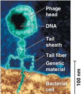
Bacteriophages (phages) are viruses that infect bacteria and consist of DNA (or RNA) enclosed in a protein coat.
Hershey and Chase (1952) used radioactive labeling to show that DNA, not protein, enters bacterial cells during infection, confirming DNA as the genetic material.
Chargaff’s Rules
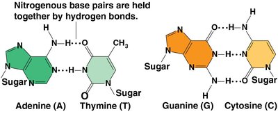
Erwin Chargaff found that DNA composition varies between species, but the amount of adenine (A) always equals thymine (T), and guanine (G) equals cytosine (C).
This base pairing regularity is known as Chargaff’s rules.
DNA Structure: The Double Helix
Discovery of the Double Helix
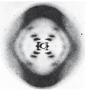
Maurice Wilkins and Rosalind Franklin used X-ray crystallography to study DNA structure, revealing its helical nature.
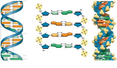
James Watson and Francis Crick built a model of DNA as a double helix with antiparallel sugar-phosphate backbones and specific base pairing.
Key Features of DNA Structure
DNA is a double helix with two antiparallel strands (5' to 3' and 3' to 5').
The backbone consists of alternating sugars (deoxyribose) and phosphates.
Nitrogenous bases (A, T, G, C) pair via hydrogen bonds: A with T, G with C.
Pairing a purine (A or G) with a pyrimidine (T or C) maintains a uniform helix width.
DNA Replication
Semiconservative Model of Replication
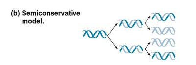
Watson and Crick proposed that each new DNA molecule consists of one parental strand and one newly synthesized strand.
This is called semiconservative replication.
Origins of Replication
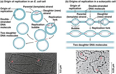
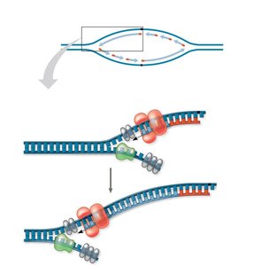
Replication begins at specific sites called origins of replication, forming replication bubbles.
Eukaryotic chromosomes have multiple origins; prokaryotes typically have one.
Enzymes and Proteins in DNA Replication
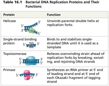
Helicase: Unwinds the DNA double helix at the replication fork.
Single-strand binding proteins: Stabilize unwound DNA strands.
Topoisomerase: Relieves overwinding strain ahead of the replication fork.
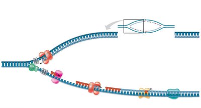
Primase: Synthesizes short RNA primers to provide a starting point for DNA synthesis.
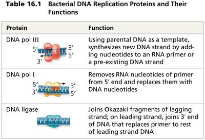
DNA polymerase III: Adds nucleotides to the 3' end of the primer, synthesizing new DNA.
DNA polymerase I: Removes RNA primers and replaces them with DNA.
DNA ligase: Joins Okazaki fragments on the lagging strand.
Protein | Function |
|---|---|
Helicase | Unwinds parental double helix at replication forks |
Single-strand binding protein | Binds to and stabilizes single-stranded DNA |
Topoisomerase | Relieves overwinding strain ahead of replication forks |
Primase | Synthesizes RNA primer |
DNA pol III | Synthesizes new DNA strand by adding nucleotides |
DNA pol I | Removes RNA primer and replaces with DNA |
DNA ligase | Joins Okazaki fragments |
Mechanism of DNA Synthesis
DNA polymerases can only add nucleotides to the 3' end; thus, DNA is synthesized in the 5' to 3' direction.
The leading strand is synthesized continuously toward the replication fork.
The lagging strand is synthesized discontinuously, forming short Okazaki fragments that are later joined by DNA ligase.
Proofreading and DNA Repair
DNA polymerases proofread each nucleotide as it is added, correcting errors.
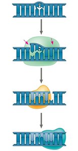
Mismatch repair enzymes fix errors missed during replication.
Nucleotide excision repair removes and replaces damaged DNA segments using nucleases, DNA polymerase, and DNA ligase.
Evolutionary Significance of DNA Mutations
Despite proofreading, some errors persist, resulting in mutations.
Mutations are the source of genetic variation, driving evolution by natural selection.
Chromosome Structure and DNA Packing
Organization of Chromosomes
Bacterial chromosomes are circular and found in the nucleoid region; eukaryotic chromosomes are linear and located in the nucleus.
In eukaryotes, DNA is combined with proteins to form chromatin.
Levels of DNA Packing
DNA wraps around histone proteins to form nucleosomes ("beads on a string").
Chromatin fibers are further folded into higher-order structures (10-nm, 30-nm, and 300-nm fibers) to fit into the nucleus.
During interphase, most chromatin is loosely packed (euchromatin), while some regions (centromeres, telomeres) are highly condensed (heterochromatin).
Regulation of Chromatin Structure
Histone proteins can be chemically modified, affecting chromatin condensation and gene expression.
The amino-terminal tails of histones play a key role in this regulation.
Summary: The molecular basis of inheritance is rooted in the structure and replication of DNA. The double helix model explains how genetic information is faithfully transmitted, while the organization of DNA into chromosomes ensures efficient packaging and regulation within the cell.
