 Back
ch 39
Study Guide - Smart Notes
 Tailored notes based on your materials, expanded with key definitions, examples, and context.
The Nervous System
Introduction
The nervous system is a complex network responsible for receiving, processing, and transmitting information throughout the body. It enables organisms to sense their environment, coordinate behavior, and maintain homeostasis. This chapter explores the cellular structure of the nervous system, the mechanisms of neural signaling, and the organization of nervous systems in animals, with a focus on the human nervous system.
Structures and Functions of Nerve Cells
Principal Cell Types
Neurons: Specialized cells that receive, process, and transmit information, and control body movements.
Glia (Glial cells): Support neurons by providing nutrients, regulating the extracellular environment, protecting against infection, repairing damage, fine-tuning communication, and speeding up electrical signal transmission.
Major Structures of a Neuron
Dendrites: Branched extensions that receive signals from other neurons or the environment.
Cell Body (Soma): Contains the nucleus and organelles; integrates incoming signals and coordinates metabolic activities.
Axon: Long projection that conducts action potentials away from the cell body toward other cells.
Synaptic Terminals: Swellings at the end of the axon that transmit information to other neurons, muscles, or glands via neurotransmitters.
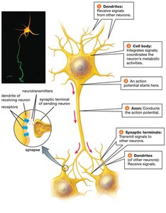
Functions of Neuron Structures
Receive information from the environment or other neurons (dendrites).
Process information and generate an electrical signal (cell body).
Conduct the electrical signal over distances (axon).
Transmit information to other cells (synaptic terminals).
Dendrites and Signal Reception
Dendrites provide a large surface area for receiving signals.
Sensory neuron dendrites respond to specific stimuli (e.g., temperature, pH, touch, odor).
Neurotransmitters released by other neurons are detected by dendrites in the brain and spinal cord.
Cell Body and Signal Integration
Signals from dendrites converge on the cell body, which sums them.
If the sum is sufficiently positive, an action potential is generated.
The cell body also performs metabolic functions and synthesizes molecules.
Axon and Signal Conduction
The axon conducts action potentials from the cell body to synaptic terminals.
Axons can be very long, forming nerves that connect the CNS to the body.
Synapses and Signal Transmission
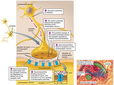
A synapse is the junction where a neuron communicates with another cell.
Neurotransmitters are released from synaptic terminals, cross the synaptic cleft, and bind to receptors on the receiving cell.
Neural Signaling: Production and Transmission of Information
Electrical Signals in Neurons
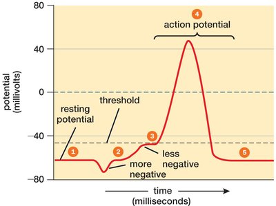
Neurons maintain a resting potential (typically −40 to −90 mV) across their plasma membrane.
Stimulation can make the membrane potential less negative; if it reaches threshold, an action potential is triggered.
Action Potentials
During an action potential, the membrane voltage rapidly rises to about +50 mV and then returns to resting potential.
Action potentials are conducted without loss of strength along the axon.
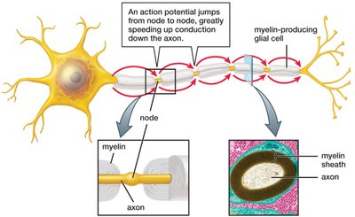
Thicker axons and myelination increase conduction speed.
Myelin and Saltatory Conduction
Myelin is a fatty insulation formed by glial cells, wrapping around axons and leaving gaps called nodes of Ranvier.
Action potentials "jump" from node to node, greatly increasing conduction speed (saltatory conduction).
Chemical Synapses
Most neuron-to-neuron communication occurs at chemical synapses via neurotransmitter release.
The synaptic cleft separates the presynaptic and postsynaptic cells.
Neurotransmitters bind to receptors, opening ion channels and generating postsynaptic potentials (PSPs).
Postsynaptic Potentials (PSPs)
Excitatory PSPs (EPSPs): Make the postsynaptic neuron more likely to fire an action potential (less negative).
Inhibitory PSPs (IPSPs): Make the neuron less likely to fire (more negative).
Integration of all PSPs at the cell body determines whether an action potential is generated.
Neurotransmitter Removal
Neurotransmitters are quickly removed from the synaptic cleft by reuptake, enzymatic breakdown, or diffusion.
Processing Information and Controlling Behavior
Neural Pathways and Behavior
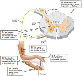
Neural pathways consist of sensory neurons, interneurons, motor neurons, and effectors (muscles/glands).
These pathways determine the type and intensity of stimuli, integrate information, and direct appropriate responses.
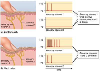
Encoding Stimulus Type and Intensity
Stimulus type is encoded by which sensory neurons and brain regions are activated.
Stimulus intensity is encoded by the frequency of action potentials and the number of neurons activated.
Integration and Output
Interneurons in the brain integrate sensory inputs and direct responses via motor neurons.
Simple behaviors (reflexes) may involve only a few neurons; complex behaviors involve extensive neural networks.
Organization of Nervous Systems
Types of Nervous Systems
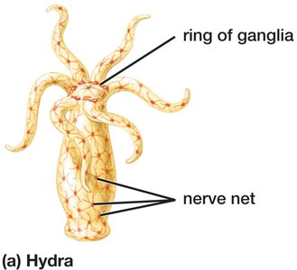
Diffuse nervous system: Found in cnidarians (e.g., Hydra), consists of a nerve net with scattered ganglia.
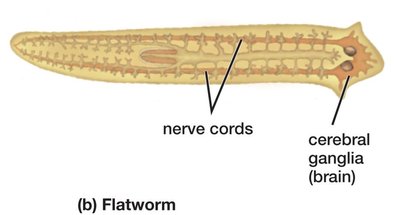
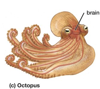
Centralized nervous system: Found in bilaterally symmetrical animals, with sense organs and ganglia concentrated in the head (cephalization).
Structures and Functions of the Human Nervous System
Central and Peripheral Nervous Systems
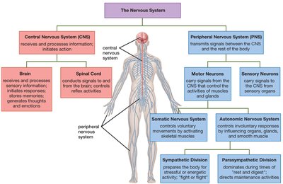
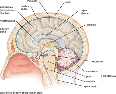
Central Nervous System (CNS): Brain and spinal cord; processes information and initiates responses.
Peripheral Nervous System (PNS): Neurons and axons outside the CNS; links the CNS to the body.
Somatic and Autonomic Nervous Systems
Somatic Nervous System: Controls voluntary movements via skeletal muscles.
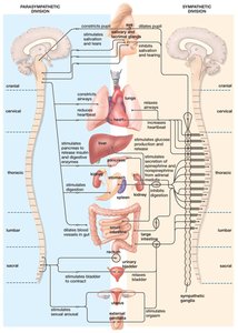
Autonomic Nervous System: Controls involuntary functions; divided into:
Sympathetic Division: Prepares the body for "fight or flight" (e.g., increases heart rate, dilates airways).
Parasympathetic Division: Promotes "rest and digest" activities (e.g., stimulates digestion, slows heart rate).
Spinal Cord Structure and Reflexes
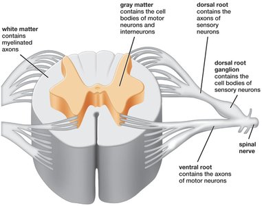
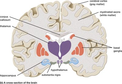
The spinal cord contains gray matter (cell bodies, interneurons) and white matter (myelinated axons).
Sensory neurons enter via dorsal roots; motor neurons exit via ventral roots.
Reflexes are rapid, involuntary responses mediated by spinal cord circuits.
Major Divisions of the Brain
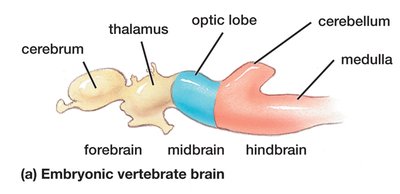
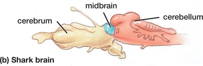
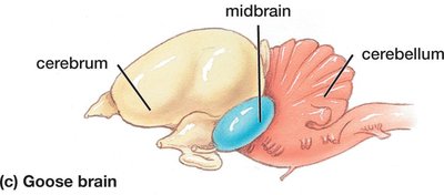
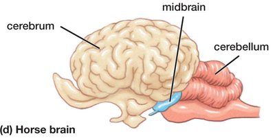
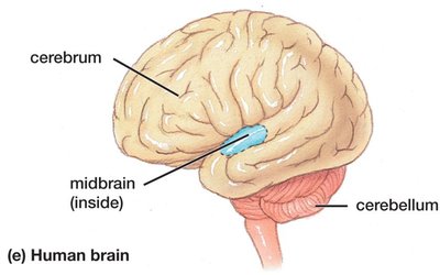
Hindbrain: Medulla, pons, cerebellum; controls automatic functions and coordinates movement.
Midbrain: Controls vision, movement, arousal, and emotion; contains dopamine-producing neurons.
Forebrain: Thalamus, hypothalamus, cerebrum; processes sensory information, regulates homeostasis, and enables higher cognitive functions.
The Cerebral Cortex
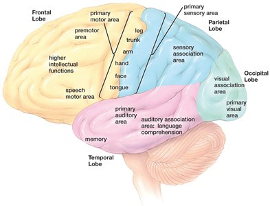
The outer layer of the cerebrum, responsible for sensory perception, voluntary movement, memory, and cognition.
Divided into four lobes: frontal, parietal, occipital, and temporal.
Contains primary sensory and motor areas, as well as association areas for interpretation and planning.
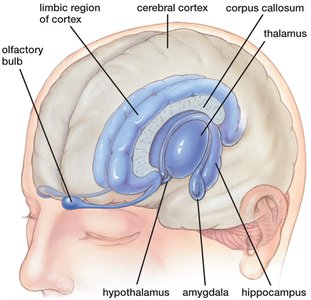
The Limbic System
A group of forebrain structures involved in emotion, memory, and homeostasis (includes hypothalamus, hippocampus, amygdala).
Generates drives (hunger, thirst), emotions, and is essential for memory formation.
Brain Lateralization
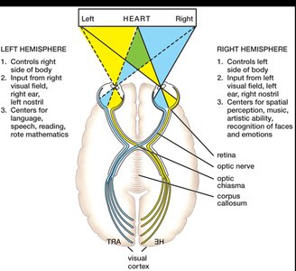
The left and right hemispheres of the brain are specialized for different functions.
Left: Language, speech, reading, rote mathematics.
Right: Musical skills, artistic ability, spatial visualization, facial recognition, emotion.
Most sensory and motor pathways cross over, so each hemisphere controls the opposite side of the body.
Learning and Memory
Short-term memory involves repeated neural activity in specific circuits (frontal/parietal lobes, hippocampus, basal ganglia).
Long-term memory involves structural changes, such as new synaptic connections or strengthening of existing ones.
The hippocampus is crucial for converting short-term to long-term memory; the cerebellum and basal ganglia are important for habits and skills.
Major Neurotransmitters
Neurotransmitter | Location | Major Functions |
|---|---|---|
Acetylcholine | Motor neuron-to-muscle synapses; autonomic nervous system; many brain areas | Activates skeletal muscles; activates target organs of the parasympathetic nervous system |
Dopamine | Midbrain | Emotion, rewards, control of movement |
Norepinephrine (noradrenaline) | Sympathetic nervous system | Activates target organs of the sympathetic nervous system |
Serotonin | Midbrain, pons, medulla | Influences mood and sleep |
Glutamate | Many brain and spinal cord areas | Major excitatory neurotransmitter in CNS |
Glycine | Spinal cord | Major inhibitory neurotransmitter in spinal cord |
GABA (gamma aminobutyric acid) | Many brain and spinal cord areas | Major inhibitory neurotransmitter in brain |
Endorphins | Many brain and spinal cord areas | Influence mood, reduce pain sensations |
Nitric oxide | Many brain areas | Important in forming memories |
Additional info: The above content integrates and expands upon the provided lecture slides, adding definitions, examples, and academic context for clarity and completeness. Images are included only when directly relevant to the explanation.
