 Back
Chapter 4: A Tour of the Cell – Study Notes
Study Guide - Smart Notes
 Tailored notes based on your materials, expanded with key definitions, examples, and context.
Chapter 4: A Tour of the Cell
Introduction to Cells
Cells are the fundamental units of life, representing the smallest structures capable of performing all activities required for life. Understanding cells is essential for studying biology, as all organisms are composed of one or more cells. The study of cells involves microscopy and biochemistry to reveal their structure and function.
Microscopy and Cell Study
Microscope Parts and Types
Microscopes are essential tools for studying cells. They magnify small objects, allowing scientists to observe cell structures that are otherwise invisible to the naked eye. There are several types of microscopes, each with specific uses:
Light Microscopes (LMs): Use visible light to illuminate specimens; suitable for living cells and general cell structure.
Electron Microscopes (EMs): Use electron beams for much higher resolution; used to study subcellular structures.
Transmission Electron Microscopes (TEMs): Provide detailed images of internal cell structures.
Scanning Electron Microscopes (SEMs): Show surface details of cells and tissues.
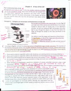
Key microscope parts: Ocular lens, objective lens, stage, coarse and fine focus knobs, light source, and base.
Magnification is the ratio of an object's image size to its real size, while resolution is the measure of clarity or the minimum distance two points can be separated and still be distinguished.
Cell Fractionation
Studying Cell Structure and Function
Cell fractionation is a technique used to separate cellular components based on size and density, allowing for the study of individual organelles and their functions. This process involves homogenizing cells and then centrifuging the mixture at various speeds to isolate different organelles.
Homogenization: Breaking open cells to release their contents.
Centrifugation: Spinning the homogenate to separate organelles by size and density.
Example: Nuclei are typically the first organelles to pellet at low speeds, followed by mitochondria, lysosomes, and ribosomes at higher speeds.
Prokaryotic vs. Eukaryotic Cells
Cell Types and Internal Membranes
Cells are classified as either prokaryotic or eukaryotic based on the presence or absence of a nucleus and membrane-bound organelles.
Prokaryotic Cells: Lack a nucleus; DNA is located in the nucleoid region. Examples include bacteria and archaea.
Eukaryotic Cells: Have a true nucleus enclosed by a nuclear envelope and possess various membrane-bound organelles. Examples include plant, animal, fungal, and protist cells.
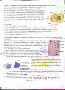
Internal membranes in eukaryotic cells compartmentalize functions, allowing for specialized environments and processes within the cell.
The Nucleus and Ribosomes
Genetic Control Centers
The nucleus contains most of the cell's genetic material and is surrounded by a double membrane called the nuclear envelope. The nucleolus within the nucleus is the site of ribosomal RNA (rRNA) synthesis and ribosome assembly.
Nuclear Envelope: Double membrane with pores for molecular transport.
Chromatin: DNA and proteins that condense to form chromosomes during cell division.
Nucleolus: Site of rRNA synthesis and ribosome assembly.
Ribosomes are complexes of rRNA and protein that carry out protein synthesis. They can be free in the cytosol or bound to the endoplasmic reticulum (ER).
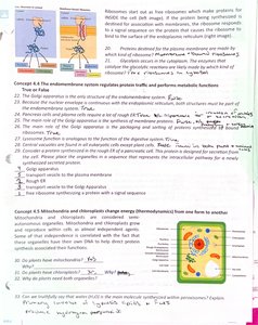
The Endomembrane System
Protein Trafficking and Metabolic Functions
The endomembrane system is a network of membranes inside and around a eukaryotic cell, involved in protein and lipid synthesis, transport, and metabolism. Major components include:
Endoplasmic Reticulum (ER): Rough ER (with ribosomes) synthesizes proteins; Smooth ER synthesizes lipids and detoxifies chemicals.
Golgi Apparatus: Modifies, sorts, and packages proteins and lipids for secretion or delivery to other organelles.
Lysosomes: Contain hydrolytic enzymes for intracellular digestion.
Vesicles and Vacuoles: Storage and transport of substances within the cell.
Example: Proteins synthesized in the rough ER are transported to the Golgi apparatus for modification and then sent to their final destinations.
Mitochondria and Chloroplasts
Energy Conversion Organelles
Mitochondria and chloroplasts are organelles that convert energy from one form to another. Mitochondria are the sites of cellular respiration, generating ATP from sugars and fats. Chloroplasts, found in plants and algae, are the sites of photosynthesis, converting solar energy into chemical energy.
Mitochondria: Double-membraned; contain their own DNA and ribosomes; site of ATP production.
Chloroplasts: Contain chlorophyll; have thylakoid membranes for light absorption and energy conversion.
Equation for Cellular Respiration:
Equation for Photosynthesis:
The Cytoskeleton
Structural Support and Movement
The cytoskeleton is a network of protein fibers that organizes structures and activities in the cell. It provides mechanical support, maintains cell shape, and facilitates cell movement and intracellular transport.
Microtubules: Hollow rods that shape the cell, guide organelle movement, and separate chromosomes during cell division.
Microfilaments (Actin Filaments): Thin rods that support the cell's shape and are involved in muscle contraction and cell motility.
Intermediate Filaments: Provide mechanical support for the cell and help anchor organelles.
Example: Cilia and flagella are structures composed of microtubules that enable cell movement.
Extracellular Components and Cell Junctions
Cell Communication and Coordination
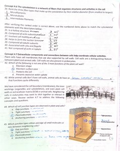
Cells interact with their environment and each other through extracellular structures and specialized junctions. The extracellular matrix (ECM) in animal cells provides structural support and mediates cell signaling. Plant cells have cell walls made of cellulose.
Tight Junctions: Prevent leakage of extracellular fluid between cells.
Desmosomes: Anchor cells together into strong sheets.
Gap Junctions: Allow ions and small molecules to pass between cells for communication.
Example: Gap junctions in heart muscle cells allow for coordinated contraction.
