 Back
Chapter 49: Nervous Systems – Structure and Function
Study Guide - Smart Notes
 Tailored notes based on your materials, expanded with key definitions, examples, and context.
Overview of Nervous Systems
Introduction to Nervous Systems
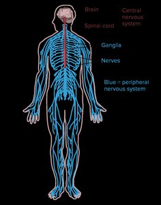
The nervous system is a complex network of specialized cells called neurons that transmit electrical and chemical signals throughout the body. It is divided into the central nervous system (CNS) and the peripheral nervous system (PNS), each with distinct structures and functions.
Central Nervous System (CNS): Consists of the brain and spinal cord; processes information and coordinates activity.
Peripheral Nervous System (PNS): Composed of nerves and ganglia outside the CNS; transmits signals between the CNS and the rest of the body.
Neurons: Structure and Function
Specialized Structure of Neurons
Neurons are the fundamental units of the nervous system, specialized for the reception, conduction, and transmission of electrical signals.
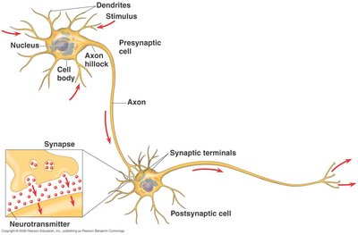
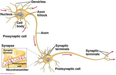
Cell Body (Soma): Contains the nucleus and most organelles.
Dendrites: Branched extensions that receive signals from other neurons.
Axon: A long projection that transmits signals to other cells; may be over 1 meter in length.
Synapse: Junction where the axon terminal of one neuron communicates with another cell via neurotransmitters.
Neuronal Signaling and Synapses
Neurons communicate through electrical impulses and chemical signals at synapses. The action potential is an all-or-nothing electrical event that travels along the axon, leading to neurotransmitter release at the synapse.
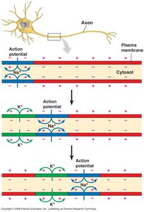
Action Potential: A rapid change in membrane potential that propagates along the axon.
Synaptic Transmission: Neurotransmitters released from the presynaptic cell bind to receptors on the postsynaptic cell, potentially triggering a new action potential.
On-Off Switch: Neurons are either at rest (negatively charged inside) or excited (positively charged inside).
Propagation of Action Potentials
Action potentials are propagated along axons by the movement of ions across the membrane. The refractory period ensures unidirectional propagation and limits the frequency of action potentials.
Refractory Period: Time during which a neuron cannot fire another action potential.
Speed of Propagation: Increased by larger axon diameter and myelination (insulation).
Evolution and Diversity of Nervous Systems
Comparative Nervous System Organization
Nervous systems vary in complexity among animal phyla, reflecting evolutionary history and lifestyle.
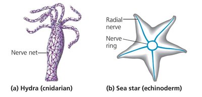
Cnidarians: Possess a diffuse nerve net.
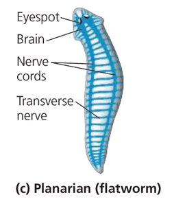
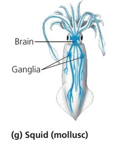
More Complex Animals: Have nerves composed of multiple neurons and centralized nervous systems.
Cephalization: Concentration of nervous tissue into a brain in bilaterally symmetrical animals.
Ganglia: Clusters of neuron cell bodies that may control specific structures.
Central and Peripheral Nervous Systems in Vertebrates
Structure and Function of the CNS
The vertebrate CNS consists of the brain and spinal cord, which process sensory information, coordinate movement, and mediate reflexes.
Brain: Processes stimuli and controls voluntary and involuntary behavior.
Spinal Cord: Transmits impulses to and from the brain; mediates reflexes.
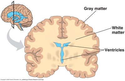
Gray Matter: Contains neuron cell bodies; located on the outside of the brain and inside the spinal cord.
White Matter: Contains myelinated axons; located inside the brain and outside the spinal cord.
Cerebrospinal Fluid: Cushions the CNS and facilitates nutrient/waste exchange.
Blood-Brain Barrier: Created by glial cells to protect the brain from harmful substances.
Peripheral Nervous System (PNS)
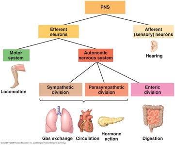
The PNS connects the CNS to the rest of the body and is divided into sensory (afferent) and motor (efferent) components.
Cranial Nerves: Connect the brain with the head and neck.
Spinal Nerves: Connect the spinal cord to the body.
Afferent Neurons: Carry sensory information to the CNS.
Efferent Neurons: Carry motor commands from the CNS to effectors.
Motor System: Controls skeletal muscles (voluntary and reflex actions).
Autonomic Nervous System: Regulates involuntary functions (smooth/cardiac muscle, glands).
Divisions of Autonomic System: Sympathetic (arousal), Parasympathetic (calming), Enteric (digestion).
Structure and Function of the Human Brain
Development and Major Regions of the Brain
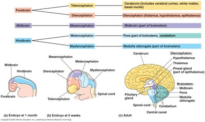
The human brain develops from three embryonic regions, each giving rise to specific adult structures with specialized functions.
Hindbrain: Includes the brainstem (pons, medulla) and cerebellum; controls homeostasis, movement, and information sharing.
Midbrain: Part of the brainstem; involved in sensory processing and alertness.
Forebrain: Includes the diencephalon (thalamus, hypothalamus, epithalamus) and cerebrum; responsible for higher-order processing, homeostasis, and circadian rhythms.
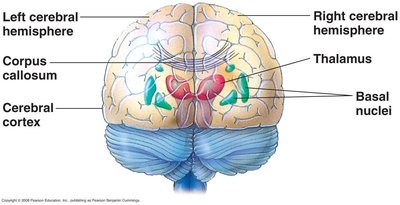
Cerebrum: Largest part of the brain; center for information processing, voluntary movement, and cognition.
Corpus Callosum: Connects the left and right cerebral hemispheres.
The Cerebral Cortex and Its Lobes
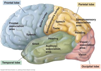
The cerebral cortex is the outer layer of the cerebrum and is responsible for conscious thought, decision-making, and sensory processing. It is divided into four lobes, each with specialized functions.
Frontal Lobe: Decision-making, planning, voluntary movement, speech production.
Parietal Lobe: Sensory perception, spatial reasoning, reading, writing.
Occipital Lobe: Visual processing.
Temporal Lobe: Hearing, language comprehension, memory.
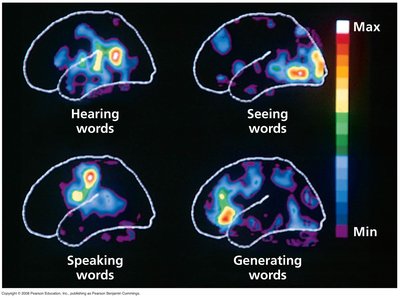
Localization of Function: Different functions are mapped to specific cortical areas (e.g., language, vision, motor control).
Lateralization, Emotion, and Consciousness
The two hemispheres of the brain are specialized for different functions, a phenomenon known as lateralization. The left hemisphere is typically associated with logic and language, while the right is involved in spatial and non-verbal tasks. Emotion and consciousness are complex and involve multiple brain regions.
Lateralization: Functional differences between the left and right hemispheres.
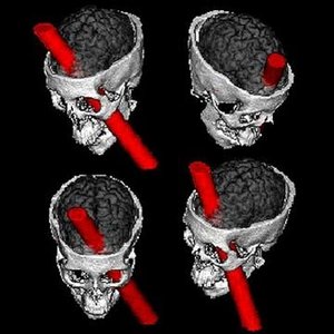
Emotion and Personality: Localized in specific brain regions; damage can alter personality (e.g., Phineas Gage case).
Neural Plasticity, Learning, and Memory
Development and Plasticity of the Nervous System
The structure of the nervous system is shaped by embryonic development, programmed cell death, and synaptic reconfiguration. Neural plasticity underlies learning and memory by altering the strength and number of synaptic connections.
Embryonic Development: Involves gene expression, signal transduction, and growth.
Neuron Die-Off: Neurons not properly connected are eliminated; about 50% are lost during development.
Synapse Reconfiguration: Excess synapses are pruned to optimize neural circuits.
Neural Plasticity: The ability of the nervous system to change in response to experience; essential for learning and memory.
Long-Term Potentiation (LTP): A process where frequent stimulation strengthens synaptic connections, making postsynaptic neurons more responsive.
Disorders of the Nervous System
Understanding Brain Function Through Disorders
Studying neurological and psychiatric disorders provides insight into normal brain function. Examples include schizophrenia, depression, drug addiction, Alzheimer's disease, and Parkinson's disease.
Schizophrenia: Affects perception and thought processes.
Depression: Involves mood regulation and neurotransmitter imbalances.
Drug Addiction: Alters reward pathways in the brain.
Alzheimer's Disease: Characterized by memory loss and cognitive decline.
Parkinson's Disease: Involves degeneration of motor control regions.
Additional info: The study of brain injuries, such as the case of Phineas Gage, has been instrumental in understanding the localization of brain functions and the biological basis of personality and behavior.
