 Back
Chapter 5: The Working Cell – Membrane Structure, Function, and Transport
Study Guide - Smart Notes
 Tailored notes based on your materials, expanded with key definitions, examples, and context.
Membrane Structure and Function
Introduction to the Plasma Membrane
The plasma membrane is a dynamic boundary that separates the interior of the cell from its external environment. It is essential for maintaining cellular integrity and regulating the movement of substances into and out of the cell. The membrane's structure and associated proteins are critical for cell survival and function.
Membrane Structure: Composed primarily of a phospholipid bilayer with embedded proteins, cholesterol, and carbohydrates.
Fluid Mosaic Model: Describes the membrane as a mosaic of protein molecules drifting in a fluid bilayer of phospholipids.
Selective Permeability: The membrane allows some substances to cross more easily than others, maintaining homeostasis.
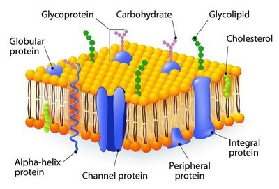
Components of the Plasma Membrane
Phospholipids: Form the basic structure, with hydrophilic heads facing outward and hydrophobic tails inward.
Proteins: Integral and peripheral proteins serve various functions such as transport, signaling, and cell recognition.
Carbohydrates: Attached to proteins (glycoproteins) or lipids (glycolipids), they play roles in cell recognition and adhesion.
Cholesterol: Stabilizes membrane fluidity across temperature changes.
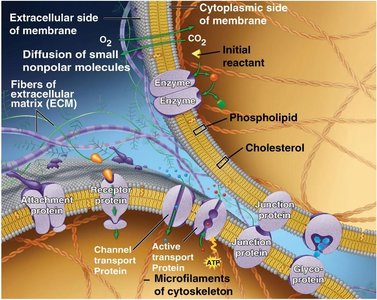
Functions of Membrane Proteins
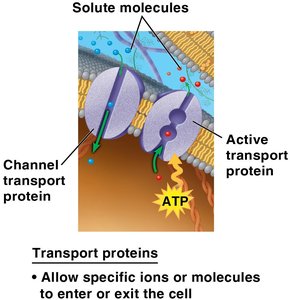
Transport: Channel and carrier proteins facilitate the movement of substances across the membrane.
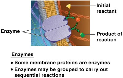
Enzymatic Activity: Some proteins act as enzymes, catalyzing reactions at the membrane surface.
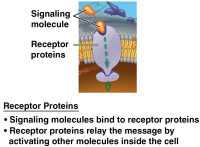
Signal Transduction: Receptor proteins transmit signals from the external environment to the cell's interior.
Cell-Cell Recognition: Glycoproteins serve as identification tags for cellular interactions.
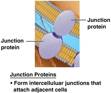
Intercellular Joining: Junction proteins connect adjacent cells.
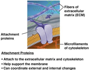
Attachment to Cytoskeleton and ECM: Provides structural support and coordinates cellular changes.
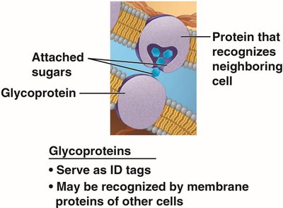
Specialized Membrane Proteins
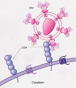
Glycoproteins: Serve as ID tags and are recognized by other cells. For example, CD4 glycoproteins on T cells are recognized by HIV.
Evolutionary Origin of Membranes
Spontaneous Formation of Membranes
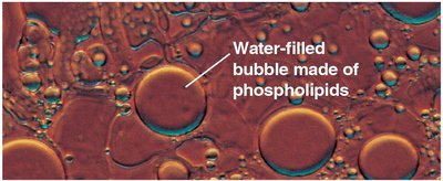
The spontaneous assembly of phospholipids into bilayers was a critical step in the origin of life. These structures formed the first cell boundaries, allowing for the compartmentalization of biological molecules.
Self-Assembly: Phospholipids naturally form bilayers in aqueous environments due to their amphipathic nature.
Requirement for Proteins: For functional cells, membranes must also contain embedded proteins to regulate transport and communication.
Membrane Transport Mechanisms
Passive Transport: Diffusion
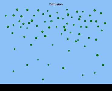
Diffusion is the movement of particles from an area of higher concentration to an area of lower concentration, driven by the random motion of molecules. This process does not require cellular energy.
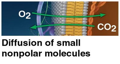
Simple Diffusion: Small, nonpolar molecules (e.g., O2, CO2) diffuse directly through the lipid bilayer.
Selective Permeability: The ease of diffusion depends on molecule size and polarity; small, uncharged molecules diffuse most readily.
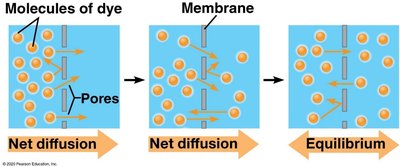
Osmosis: Diffusion of Water
Osmosis is the diffusion of water across a selectively permeable membrane. Water moves from regions of higher free water concentration (lower solute concentration) to regions of lower free water concentration (higher solute concentration).
Direction of Water Movement: Water moves toward the solution with higher solute concentration if the membrane is impermeable to the solute.
Osmotic Terms: Hypotonic (lower solute), isotonic (equal solute), hypertonic (higher solute) relative to the cell interior.
Water Balance and Tonicity
Cells must regulate water balance to survive. Tonicity describes the ability of a surrounding solution to cause a cell to gain or lose water.
Hypotonic Solution: Lower solute concentration than the cell; water enters the cell, which may burst (lysis) in animal cells or become turgid in plant cells.
Isotonic Solution: Equal solute concentration; no net water movement.
Hypertonic Solution: Higher solute concentration; water leaves the cell, causing it to shrink (crenation in animal cells, plasmolysis in plant cells).
Facilitated Diffusion
Facilitated diffusion is the passive movement of molecules across the membrane via specific transport proteins. This process does not require energy and relies on the concentration gradient.
Transport Proteins: Channel and carrier proteins enable the passage of ions and polar molecules.
Aquaporins: Specialized channel proteins that facilitate rapid water transport.
Active Transport
Active transport moves substances against their concentration gradients, requiring energy input, usually from ATP. This process is essential for maintaining concentration differences across membranes.
Mechanism: Transport proteins (pumps) use ATP to move ions or molecules from low to high concentration.
Example: The sodium-potassium pump in animal cells maintains electrochemical gradients.
Bulk Transport: Exocytosis and Endocytosis
Large molecules and particles are transported across membranes via vesicle-mediated processes.
Exocytosis: The export of large molecules (e.g., proteins, polysaccharides) by vesicle fusion with the plasma membrane.
Endocytosis: The import of large molecules by engulfing them in vesicles. Includes phagocytosis (cell eating), pinocytosis (cell drinking), and receptor-mediated endocytosis (specific uptake).
Summary Table: Types of Membrane Transport
Transport Type | Energy Required? | Direction | Example |
|---|---|---|---|
Simple Diffusion | No | High to Low | O2, CO2 |
Facilitated Diffusion | No | High to Low | Glucose, ions via channels |
Osmosis | No | High to Low (water) | Water via aquaporins |
Active Transport | Yes (ATP) | Low to High | Na+/K+ pump |
Exocytosis | Yes | Out of cell | Secretion of hormones |
Endocytosis | Yes | Into cell | Phagocytosis of bacteria |
Key Equations
Fick's Law of Diffusion:
Where J is the rate of diffusion, D is the diffusion coefficient, and \frac{dC}{dx} is the concentration gradient.
Examples and Applications
Osmoregulation in Sharks: Bull sharks can survive in both salt and fresh water by regulating solute and water balance using specialized organs.
Contractile Vacuoles in Paramecium: Expel excess water in hypotonic environments to prevent cell lysis.
HIV Recognition: CD4 glycoproteins on T cells are recognized by HIV, illustrating the importance of membrane proteins in disease processes.
Concept Check
Osmosis: To level solutions separated by a semipermeable membrane, add water to the side with higher solute concentration or add solute to the side with lower solute concentration.
Membrane Proteins: The region of a membrane protein within the membrane is hydrophobic, while the region exposed to the aqueous environment is hydrophilic.
