 Back
BackChapter 8: The Cellular Basis of Reproduction and Inheritance – Study Notes
Study Guide - Smart Notes
 Tailored notes based on your materials, expanded with key definitions, examples, and context.
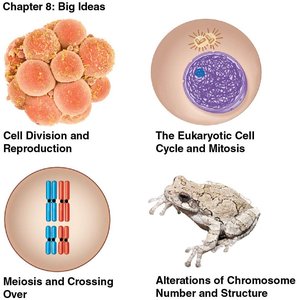
Cell Division and Reproduction
Introduction to Cell Division
Cell division is a fundamental process in all living organisms, essential for growth, development, and reproduction. It ensures the continuity of life by producing new cells from preexisting ones.
Asexual reproduction produces offspring that are genetically identical to the parent (clones).
Sexual reproduction generates offspring with unique combinations of genes, increasing genetic diversity.
Before division, the parent cell duplicates its chromosomes, which are DNA-protein complexes containing genetic information.
Examples of Reproduction
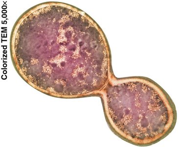
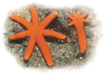
Asexual reproduction can occur via budding (e.g., yeast) or fragmentation and regeneration (e.g., starfish).
Sexual reproduction involves the fusion of gametes, resulting in genetically varied offspring.

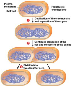
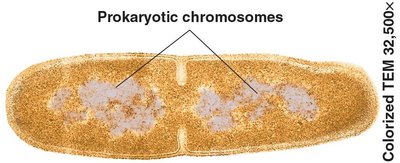
Prokaryotic Cell Division: Binary Fission
Binary Fission in Prokaryotes
Prokaryotic cells, such as bacteria, reproduce asexually by binary fission, a process that divides one cell into two genetically identical daughter cells.
Most prokaryotic genes are carried on a single, circular DNA molecule (chromosome).
During binary fission:
The chromosome is duplicated.
The copies move apart as the cell elongates.
The plasma membrane pinches inward, and a new cell wall forms, dividing the cell.
The Eukaryotic Cell Cycle and Mitosis
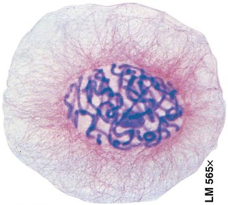
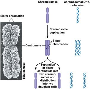
Chromosomes and Chromatin
Eukaryotic cells have multiple, linear chromosomes located in the nucleus. Each chromosome consists of a long DNA molecule and associated proteins, forming chromatin.
Chromosomes are visible only during cell division; otherwise, DNA exists as loosely packed chromatin.
Before division, chromosomes duplicate, forming sister chromatids joined at a centromere.
Human somatic cells have 46 chromosomes (23 pairs).
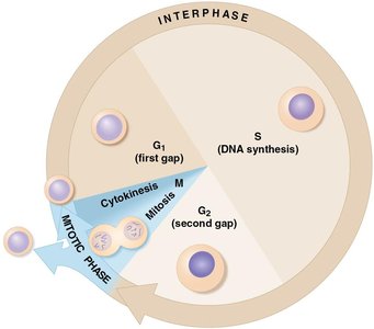
The Cell Cycle
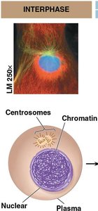
The cell cycle is an ordered sequence of events from the formation of a cell to its own division. It consists of interphase and the mitotic (M) phase.
Interphase (G1, S, G2): Cell grows, replicates DNA, and prepares for division.
Mitotic phase (M phase): Includes mitosis (nuclear division) and cytokinesis (cytoplasmic division).
Phases of Mitosis
Mitosis is the process by which duplicated chromosomes are equally distributed into two daughter nuclei. It consists of several phases:
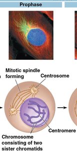
Prophase: Chromatin condenses into visible chromosomes; mitotic spindle begins to form.
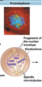
Prometaphase: Nuclear envelope fragments; spindle microtubules attach to kinetochores.
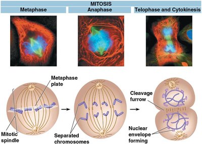
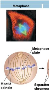
Metaphase: Chromosomes align at the metaphase plate.
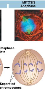
Anaphase: Sister chromatids separate and move toward opposite poles.
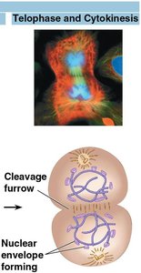
Telophase: Nuclear envelopes reform; chromosomes decondense.
Cytokinesis: Division of the cytoplasm, resulting in two daughter cells.
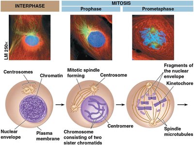
Details of Each Mitotic Phase
Interphase: Cell grows, DNA replicates, centrosomes duplicate.
Prophase: Chromosomes condense, spindle forms, centrosomes move apart.
Prometaphase: Nuclear envelope breaks down, spindle fibers attach to kinetochores.
Metaphase: Chromosomes align at the metaphase plate.
Anaphase: Centromeres split, sister chromatids move to opposite poles.
Telophase: Chromosomes decondense, nuclear envelopes reform.
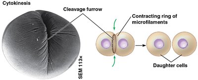
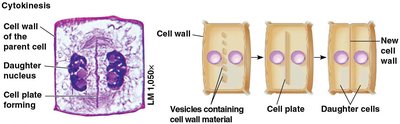
Cytokinesis: Animal cells form a cleavage furrow; plant cells form a cell plate.

Cytokinesis in Animal and Plant Cells
In animal cells, cytokinesis occurs by cleavage, forming a cleavage furrow that pinches the cell in two.
In plant cells, vesicles form a cell plate that develops into a new cell wall, dividing the cell.
Regulation of the Cell Cycle
Control of Cell Division
Cell division is regulated by environmental factors and internal signals to ensure proper growth and development.
Anchorage dependence: Cells must be attached to a surface to divide.
Density-dependent inhibition: Cells stop dividing when they become crowded.
Growth factors: Chemical signals that stimulate cell division.
Cell Cycle Checkpoints
G1 checkpoint: Determines if the cell will divide or enter a nondividing state (G0).
G2 checkpoint: Ensures DNA is undamaged before mitosis.
Mitotic checkpoint: Ensures all chromosomes are properly attached to the spindle before anaphase.
Cancer and the Cell Cycle
Cancer cells divide uncontrollably, forming tumors.
Malignant tumors can invade other tissues (metastasis).
Radiation and chemotherapy target rapidly dividing cells.
Meiosis and Crossing Over
Homologous Chromosomes and Ploidy
Somatic cells are diploid (2n), containing two sets of chromosomes (one from each parent).
Gametes (sperm and eggs) are haploid (n), containing one set of chromosomes.
Fertilization restores diploidy, forming a zygote.
Overview of Meiosis
Meiosis is a two-division process that reduces chromosome number by half, producing four genetically unique haploid cells.
Meiosis I: Homologous chromosomes separate.
Meiosis II: Sister chromatids separate (similar to mitosis).
Crossing over during prophase I increases genetic variation.
Comparison of Mitosis and Meiosis
Mitosis produces two genetically identical diploid cells for growth and repair.
Meiosis produces four genetically unique haploid gametes for sexual reproduction.
Genetic Variation
Independent assortment of chromosomes and random fertilization contribute to genetic diversity.
Crossing over during prophase I further increases variability.
Alterations of Chromosome Number and Structure
Nondisjunction and Aneuploidy
Nondisjunction: Failure of homologous chromosomes or sister chromatids to separate during meiosis, leading to abnormal chromosome numbers (aneuploidy).
Example: Trisomy 21 (Down syndrome) results from an extra chromosome 21.
Abnormal numbers of sex chromosomes can lead to syndromes such as Klinefelter (XXY) or Turner (XO).
Karyotyping
A karyotype is a photographic inventory of an individual's chromosomes, used to detect chromosomal abnormalities.
Chromosomal Structural Changes
Chromosome breakage can cause deletions, duplications, inversions, or translocations, leading to genetic disorders or cancer.
Summary Table: Mitosis vs. Meiosis
Feature | Mitosis | Meiosis |
|---|---|---|
Number of chromosomal duplications | 1 | 1 |
Number of cell divisions | 1 | 2 |
Number of daughter cells produced | 2 | 4 |
Number of chromosomes in daughter cells | Same as parent (diploid) | Half of parent (haploid) |
How chromosomes line up during metaphase | Single file | Homologous pairs (Meiosis I), single file (Meiosis II) |
Genetic relationship of daughter cells to parent | Identical | Unique |
Functions in the human body | Growth, repair, asexual reproduction | Production of gametes |
Key Terms and Concepts
Chromosome: DNA-protein structure carrying genetic information.
Sister chromatids: Identical copies of a duplicated chromosome, joined at the centromere.
Centromere: Region where sister chromatids are attached.
Homologous chromosomes: Chromosome pairs with genes for the same traits.
Diploid (2n): Two sets of chromosomes.
Haploid (n): One set of chromosomes.
Gamete: Haploid reproductive cell (sperm or egg).
Zygote: Diploid cell formed by fertilization.
Nondisjunction: Failure of chromosomes to separate properly during meiosis.
Karyotype: Ordered display of an individual's chromosomes.
