 Back
BackChromosomal Abnormalities and Genetic Disorders: Structure, Number, and Detection
Study Guide - Smart Notes
 Tailored notes based on your materials, expanded with key definitions, examples, and context.
Chromosomes and the Karyotype
Chromosome Structure and Function
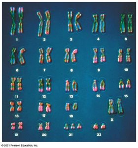
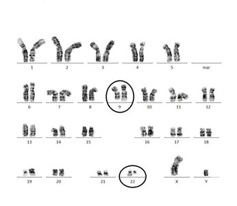
Chromosomes are thread-like structures composed of DNA and proteins, carrying genetic information essential for cellular function and inheritance. The karyotype is a visual representation of the complete set of chromosomes in a cell, used to detect abnormalities in chromosome number and structure.
Karyotype: An ordered display of chromosomes from a cell, typically arranged by size and shape.
Applications: Used for prenatal screening, detection of chromosomal abnormalities, and determination of sex.
Example: Human karyotypes typically show 46 chromosomes, including 22 pairs of autosomes and 1 pair of sex chromosomes.
Chromosome Alterations
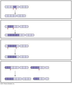
Alterations in chromosome structure can lead to genetic disorders. Four main types of structural changes are deletion, duplication, inversion, and translocation.
Deletion: Removal of a chromosomal segment.
Duplication: Repetition of a segment.
Inversion: Reversal of a segment within a chromosome.
Translocation: Movement of a segment to a nonhomologous chromosome.
Large Scale Chromosome Changes
Aneuploidy: Abnormal Chromosome Number
Aneuploidy refers to the presence of an abnormal number of chromosomes in a cell. This condition can disrupt genetic balance and lead to syndromes with characteristic symptoms.
Trisomy: Presence of an extra chromosome (e.g., trisomy 21 in Down syndrome).
Monosomy: Absence of a chromosome (e.g., monosomy X in Turner syndrome).
Example: Down syndrome (trisomy 21) results from three copies of chromosome 21.
Common Aneuploidy Syndromes
Down Syndrome (Trisomy 21): Characterized by intellectual disability, distinctive facial features, and increased risk of heart defects.
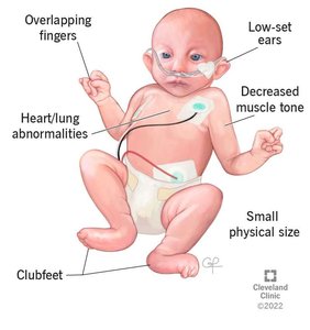
Edwards Syndrome (Trisomy 18): Severe intellectual disabilities, physical abnormalities, most affected infants die before or shortly after birth.
Patau Syndrome (Trisomy 13): Profound developmental delays, heart defects, brain abnormalities, high infant mortality.
Aneuploidy of Sex Chromosomes
Trisomy X (Triple X Syndrome): XXX females, usually healthy and fertile.
Turner Syndrome (Monosomy X): X0 females, sterile, normal intelligence, may lack secondary sexual characteristics without hormone therapy.
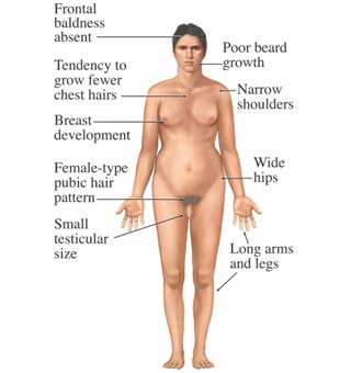
Klinefelter Syndrome (XXY): Males with extra X chromosome, sterile, characteristic physical features.
XYY Syndrome: Males with extra Y chromosome, few symptoms, often taller than average.
X Inactivation in Female Mammals
Gene Dosage and Barr Body Formation
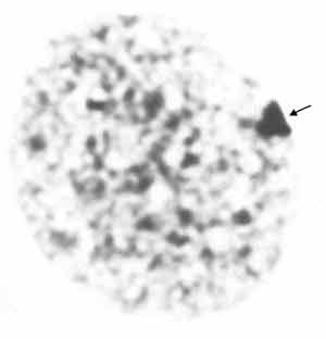
In female mammals, one of the two X chromosomes in each cell is randomly inactivated during embryonic development to balance gene dosage. The inactive X chromosome condenses into a structure called a Barr body.
Barr Body: Inactive X chromosome visible in the nucleus.
Gene Dosage: Ensures females do not have double the expression of X-linked genes compared to males.
Alterations of Chromosome Structure
Types of Structural Changes
Chromosome breakage can lead to four main types of structural changes: deletion, duplication, inversion, and translocation. These changes can disrupt gene function and lead to genetic disorders.
Deletion: Loss of genetic material.
Duplication: Extra copies of genetic material.
Inversion: Reversed orientation of genetic material.
Translocation: Exchange of segments between nonhomologous chromosomes.
Translocation and Genetic Disorders
Translocation can result in genetic disorders such as certain cases of Down syndrome and the Philadelphia chromosome in chronic myelogenous leukemia (CML).
Translocation Down Syndrome: Part of chromosome 21 attaches to another chromosome, resulting in three copies of chromosome 21 genes.
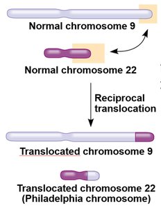
Philadelphia Chromosome: Reciprocal translocation between chromosomes 9 and 22, associated with CML.
Fetal Testing and Detection of Chromosomal Abnormalities
Methods of Prenatal Screening
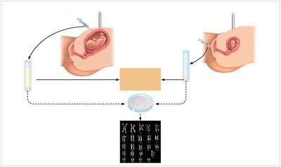
Several methods are used to detect chromosomal abnormalities in fetuses, including amniocentesis, chorionic villus sampling (CVS), and noninvasive prenatal testing (NIPT).
Amniocentesis: Removal and testing of amniotic fluid.
Chorionic Villus Sampling (CVS): Removal and testing of placental tissue.
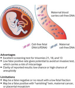
Noninvasive Prenatal Testing (NIPT): Analysis of fetal DNA from maternal blood.
Summary Table: Chromosomal Abnormalities
Type | Chromosome(s) Affected | Symptoms | Viability |
|---|---|---|---|
Down Syndrome | Trisomy 21 | Intellectual disability, heart defects, facial features | Survives to adulthood |
Edwards Syndrome | Trisomy 18 | Severe disabilities, physical abnormalities | Most die before or shortly after birth |
Patau Syndrome | Trisomy 13 | Developmental delays, organ defects | Most die within first year |
Turner Syndrome | Monosomy X | Sterile, normal intelligence | Viable |
Klinefelter Syndrome | XXY | Sterile, physical features | Viable |
Triple X Syndrome | XXX | Healthy, fertile | Viable |
XYY Syndrome | XYY | Taller, few symptoms | Viable |
Key Equations and Concepts
Aneuploidy: (where is the haploid number)
Karyotype Analysis: Used to detect chromosomal abnormalities
Review Questions
For what purpose(s) might a karyotype be prepared?
A. For prenatal screening, to determine if a fetus has the correct number of chromosomes
B. To detect the possible presence of chromosomal abnormalities such as deletions, inversions, or translocations
C. To determine whether a fetus is male or female
D. The first and second answers are correct.
E. The first three answers are correct.
Additional info: Karyotyping and chromosomal analysis are essential tools in genetics for diagnosing and understanding chromosomal disorders, gene dosage effects, and inheritance patterns.
