 Back
BackChromosome Packing and Mutations: Structure, Function, and Genetic Variation
Study Guide - Smart Notes
 Tailored notes based on your materials, expanded with key definitions, examples, and context.
Chromosome Structure and DNA Packaging
Hierarchy of Genetic Material
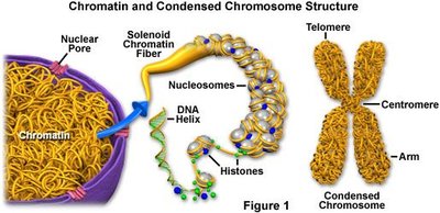
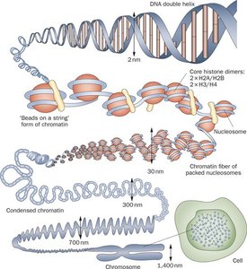
The genetic material in eukaryotic cells is organized in a hierarchical manner, from the smallest nucleotide to the entire cell. Understanding this hierarchy is essential for grasping how genetic information is stored and expressed.
Nucleotide (base): The basic unit of DNA, consisting of a sugar, phosphate, and nitrogenous base.
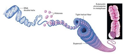
Double helix: The structure formed by two strands of DNA coiled around each other.
Chromatin: DNA wrapped around histone proteins, forming a complex that can be further packed.
Chromosome: Highly condensed chromatin, visible during cell division.
Nucleus: The organelle containing chromosomes.
Cell: The basic unit of life, containing the nucleus.
Size Comparison: Chromosomes are approximately 700 times wider than the DNA double helix, with the double helix measuring about 2 nm across and chromosomes about 1400 nm across.

Karyotypes and Chromosome Number
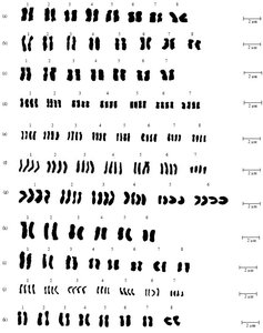
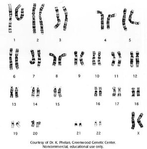
What Can Be Seen in Karyotypes?
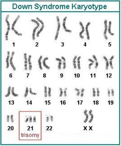
Karyotyping is a technique used to visualize chromosomes in a cell, allowing for the detection of abnormalities in chromosome number and structure.
Abnormal chromosome numbers: Such as trisomy 21 (Down syndrome).
Sex chromosomes: Identification of X and Y chromosomes.
Ploidy level: The number of sets of chromosomes in a cell.
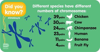
Species differences: Different species have characteristic chromosome numbers.

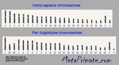
What Cannot Be Seen in Karyotypes?
Karyotypes do not reveal information about individual gene sequences, alleles, or single nucleotide differences. For such details, DNA must be analyzed at the double helix level.
DNA Mutations
Types of Mutations
Mutations are changes in genetic information and can occur at different levels:
Chromosomal Structure Changes: Involve large segments of chromosomes, such as translocations, deletions, duplications, or inversions.
DNA Mutations: Affect single bases (point mutations) or involve insertions/deletions (indels).
Chromosomal Structure Changes: Translocation Example
Translocation is a chromosomal abnormality where a segment of one chromosome is transferred to another. The Philadelphia chromosome, resulting from a translocation between chromosomes 9 and 22, is implicated in chronic myeloid leukemia (CML).

DNA Mutations: Point Mutations
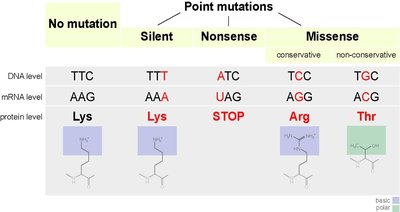
Point mutations involve the substitution of a single nucleotide. These are also known as single nucleotide polymorphisms (SNPs) and can be classified as:
Silent: No change in the amino acid sequence.
Nonsense: Results in a premature stop codon.
Missense: Changes one amino acid to another.
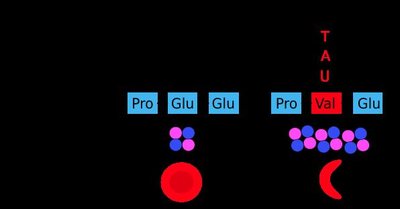
Example: Sickle Cell Disease
Sickle cell disease is caused by a missense mutation in the hemoglobin gene, where a single nucleotide change results in the substitution of valine for glutamic acid.
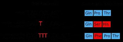
DNA Mutations: Indels and Frameshift Mutations
Indels are insertions or deletions of bases in DNA. If not in multiples of three, they cause a frameshift mutation, altering the reading frame and often resulting in a non-functional protein.

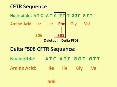
Example: Cystic Fibrosis Mutation
The most common mutation causing cystic fibrosis is the deletion of three nucleotides, resulting in the loss of phenylalanine at position 508 in the CFTR protein.
Causes of Mutations
Mutation Origins
Mutations can arise from errors during DNA replication, mistakes in crossing over during meiosis, or exposure to mutagenic chemicals and UV radiation.
Replication errors: Spontaneous mistakes during DNA synthesis.
Crossing over errors: Incorrect exchange of genetic material during meiosis.
Mutagens: Chemicals or radiation that increase mutation rates.
Inbreeding and Mutation Expression
Inbreeding does not cause new mutations but increases the likelihood of expressing recessive alleles already present in the population. When closely related individuals mate, offspring are more likely to inherit two copies of harmful recessive alleles, leading to the expression of negative phenotypes.
Example: Canine hip dysplasia is more common in inbred populations due to increased homozygosity of recessive alleles.
Summary Table: Types of DNA Mutations
Mutation Type | Description | Effect | Example |
|---|---|---|---|
Point Mutation | Single base substitution | Silent, nonsense, or missense | Sickle cell disease |
Indel | Insertion or deletion of bases | Frameshift if not in multiples of three | Cystic fibrosis (Delta F508) |
Chromosomal Structure Change | Large segment rearrangement | Translocation, deletion, duplication, inversion | Philadelphia chromosome (CML) |
Mutation Review and Additional Resources
For further review, students are encouraged to watch educational videos such as those from the Amoeba Sisters and Khan Academy, which provide visual explanations of mutation types and their effects.
Additional info: The notes expand on the original content by providing definitions, examples, and academic context for each mutation type and chromosome structure, ensuring completeness and clarity for exam preparation.
