 Back
BackChromosome Structure and Chromatin Organization
Study Guide - Smart Notes
 Tailored notes based on your materials, expanded with key definitions, examples, and context.
Chromosome Structure and Chromatin Organization
Characteristics of Eukaryotic Cells and DNA
Eukaryotic cells are defined by the presence of a nucleus, which houses the genetic material in the form of chromosomes. The human genome contains approximately 3 billion base pairs, encoding all proteins and genetic traits. In eukaryotes, DNA is divided into long, double-stranded molecules called chromosomes.
Nucleus: Membrane-bound organelle containing chromosomes.
Chromosomes: Linear DNA molecules with associated proteins, unique to each species.
Genome Size: Human haploid genome ~3.2 billion base pairs; total DNA length ~2.2 meters per cell.
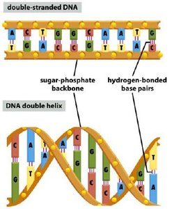
DNA Compaction and Chromosome Packaging
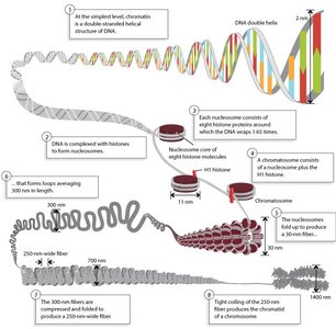
Given the vast length of DNA, it must be highly compacted to fit within the small nuclear volume (5–10 μm in diameter). This compaction is achieved through hierarchical packaging into chromatin and higher-order structures.
DNA Compaction: DNA is wrapped around histone proteins to form nucleosomes, further folded into higher-order fibers and ultimately chromosomes.
Chromatin: The complex of DNA and proteins (mainly histones) that forms chromosomes.
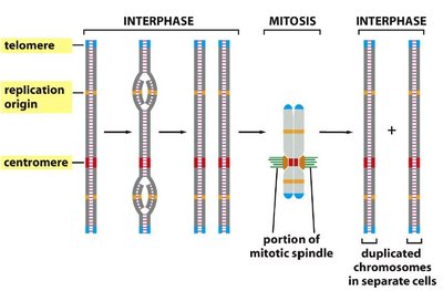
Chromosome Structure and Key Features
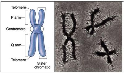
Chromosomes have distinct structural regions essential for their function and inheritance during cell division.
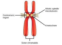
Chromatid: One of two identical copies of a replicated chromosome, joined at the centromere.
Centromere: Region where sister chromatids are held together and kinetochore forms.
Kinetochore: Protein complex at the centromere, essential for chromosome movement during mitosis.
Telomeres: Repetitive DNA sequences at chromosome ends, protecting them from degradation.
Chromosome Compaction During the Cell Cycle
The degree of chromosome compaction varies throughout the cell cycle. Chromosomes are most condensed during mitosis (metaphase), while in interphase, they are less compact and more accessible for gene expression.
Metaphase Chromosomes: Highly condensed, visible under a microscope.
Interphase Chromosomes: Less condensed, variable compaction depending on activity.
Euchromatin and Heterochromatin
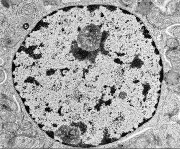
Chromatin exists in two main forms during interphase, each with distinct structural and functional properties:
Euchromatin: Loosely packed, light-staining, transcriptionally active regions, often found in the nuclear interior.
Heterochromatin: Densely packed, dark-staining, transcriptionally inactive regions, often located at the nuclear periphery. Includes centromeres and telomeres with repetitive DNA.
Nucleosome Structure and DNA Packaging
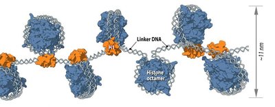
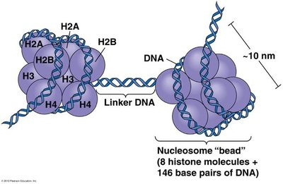
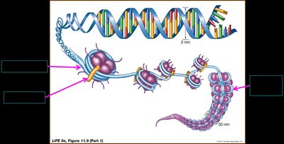
The nucleosome is the fundamental unit of chromatin, consisting of DNA wrapped around a histone octamer. This structure compacts DNA approximately sevenfold and serves as the first level of DNA organization in the nucleus.
Nucleosome: 146–147 base pairs of DNA wrapped around an octamer of histone proteins (2x H2A, H2B, H3, H4).
Histone H1: Involved in higher-order chromatin structure and further compaction.
Beads-on-a-string: Appearance of nucleosomes under electron microscopy.
Higher-Order Chromatin Structure
Chromatin undergoes further folding and organization into higher-order structures, ultimately forming the highly condensed metaphase chromosome. This process involves additional proteins and structural motifs.
30-nm Fiber: Nucleosomes are further coiled into a 30-nm fiber, stabilized by histone H1.
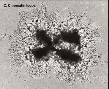
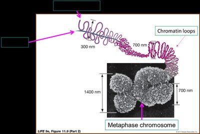
Chromatin Loops: 30-nm fibers form loops attached to a protein scaffold.
Metaphase Chromosome: The most condensed form, visible during cell division.
Chromosome Territories and Nuclear Organization
During interphase, individual chromosomes occupy distinct regions within the nucleus, known as chromosome territories. This spatial organization limits intermingling and influences gene regulation.
Chromosome Territories: Discrete nuclear regions occupied by individual chromosomes.
Genomic Proximity: DNA-DNA contacts occur predominantly within the same chromosome.
Regulation of Gene Expression by Chromatin Remodeling
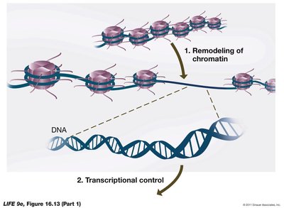
Gene expression in eukaryotes is tightly regulated at multiple levels, including chromatin structure. Chromatin remodeling alters DNA accessibility, enabling or restricting transcription factor binding and gene activation.
Chromatin Remodeling: Dynamic modification of chromatin structure to regulate DNA accessibility.
Histone Modifications: Covalent modifications (e.g., acetylation, methylation) of histone tails alter chromatin structure and gene expression.
ATP-dependent Remodelers: Protein complexes that reposition or restructure nucleosomes using ATP hydrolysis.
Histone Acetylation and Chromatin Accessibility
Histone acetyltransferases (HATs) add acetyl groups to lysine residues on histone tails, neutralizing their positive charge and loosening DNA-histone interactions. This process increases DNA accessibility and promotes transcription. Histone deacetylases (HDACs) remove acetyl groups, leading to chromatin condensation and gene repression.
HATs: Enzymes that acetylate histone tails, activating transcription.
HDACs: Enzymes that remove acetyl groups, repressing transcription.
Clinical Relevance: HDAC inhibitors are explored as cancer therapies to reactivate silenced tumor suppressor genes.
Mechanisms of Chromatin Remodeling
Chromatin remodeling complexes use several mechanisms to increase local DNA accessibility:
Nucleosome sliding
Nucleosome displacement
Partial histone displacement
Replacement of histone subunits with variants
Summary Table: Key Chromosome and Chromatin Terms
Term | Definition |
|---|---|
Nucleus | Membrane-bound organelle containing chromosomes |
Chromosome | Linear DNA molecule with associated proteins |
Chromatin | Complex of DNA and proteins forming chromosomes |
Chromatid | One of two identical copies of a replicated chromosome |
Centromere | Region joining sister chromatids; kinetochore forms here |
Kinetochore | Protein complex for chromosome movement in mitosis |
Telomere | Repetitive DNA at chromosome ends |
Euchromatin | Loosely packed, transcriptionally active chromatin |
Heterochromatin | Densely packed, transcriptionally inactive chromatin |
Nucleosome | DNA wrapped around histone octamer |
Histone | Protein component of nucleosomes |
Chromatin Remodeling | Dynamic modification of chromatin structure |
HAT | Histone acetyltransferase, activates transcription |
HDAC | Histone deacetylase, represses transcription |
