 Back
BackChromosome Structure and DNA Packing in Eukaryotes
Study Guide - Smart Notes
 Tailored notes based on your materials, expanded with key definitions, examples, and context.
Chromosome Structure and DNA Packing
Overview of Chromosome Organization
Chromosomes are highly organized structures that package and manage the long DNA molecules found in cells. The organization of DNA into chromosomes is essential for the storage, expression, and transmission of genetic information. The structure and packing of chromosomes differ between prokaryotes and eukaryotes, with eukaryotic chromosomes displaying complex levels of organization involving both DNA and proteins.
Bacterial Chromosomes: Typically consist of a single, circular DNA molecule associated with specific proteins. The DNA is supercoiled and localized in a region called the nucleoid, which is not membrane-bound.
Eukaryotic Chromosomes: Composed of linear DNA molecules associated with a large number of proteins, forming a complex called chromatin. Each human chromosome contains a single DNA molecule that, if stretched out, would be several centimeters long.
DNA Packing in Eukaryotes
To fit within the nucleus, eukaryotic DNA undergoes multiple levels of packing, resulting in the formation of chromatin. This packing is dynamic and changes throughout the cell cycle.
Chromatin: The combination of DNA and proteins that makes up chromosomes. Chromatin can exist in more or less condensed forms depending on the cell's needs.
Levels of Packing: DNA is wrapped around histone proteins to form nucleosomes, which are further folded and organized into higher-order structures.
Levels of Chromatin Organization
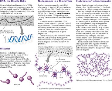
DNA, Histones, and Nucleosomes
The basic unit of DNA packing in eukaryotic cells is the nucleosome. Nucleosomes are formed when DNA wraps around histone protein cores, creating a "beads-on-a-string" structure known as the 10-nm fiber.
Histones: Small, positively charged proteins that bind to negatively charged DNA, facilitating its compaction. There are five main types of histones: H1, H2A, H2B, H3, and H4.
Nucleosome: Consists of DNA wrapped around a histone octamer (two each of H2A, H2B, H3, and H4). The nucleosome core particle is about 10 nm in diameter.
Linker DNA: The stretch of DNA between nucleosomes, which can be bound by histone H1 to further stabilize the structure.
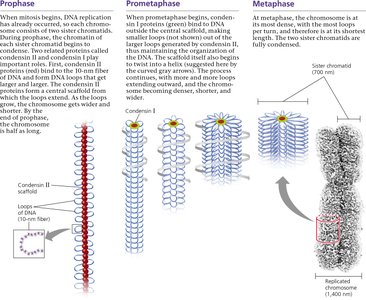
Higher-Order Chromatin Structure
Nucleosomes are further organized into more compact structures through additional folding and looping, ultimately forming the highly condensed chromosomes seen during cell division.
30-nm Fiber: Nucleosomes coil to form a thicker fiber, stabilized by interactions between histone tails and linker DNA.
Looped Domains: The 30-nm fiber forms loops attached to a protein scaffold, further compacting the DNA.
Metaphase Chromosome: During mitosis, chromatin becomes highly condensed, forming the characteristic metaphase chromosome structure.
Chromatin States: Euchromatin and Heterochromatin
Euchromatin vs. Heterochromatin
Chromatin can exist in two main states, which differ in their degree of compaction and functional properties:
Euchromatin: Loosely packed chromatin that is accessible to the transcription machinery, allowing gene expression. It appears as a diffuse mass in the nucleus during interphase.
Heterochromatin: Densely packed chromatin, often found at centromeres and telomeres, that is generally transcriptionally inactive due to its inaccessibility. It appears as irregular clumps under the microscope.
Table: Comparison of Euchromatin and Heterochromatin
Property | Euchromatin | Heterochromatin |
|---|---|---|
Compaction | Less condensed | Highly condensed |
Transcriptional Activity | Active (genes accessible) | Inactive (genes inaccessible) |
Location | Throughout nucleus | Centromeres, telomeres, specific regions |
Dynamic Nature of Chromatin
Chromatin structure is not static; it is dynamically modified during the cell cycle and in response to cellular signals. Chemical modifications of histones (such as acetylation, methylation, and phosphorylation) can alter chromatin structure and regulate gene expression.
During interphase, chromatin is generally less condensed, allowing access for DNA replication and transcription.
As the cell prepares for mitosis, chromatin condenses to form visible chromosomes, ensuring accurate segregation of genetic material.
Key Concepts and Review Questions
Nucleosome Structure: A nucleosome consists of DNA wrapped around a histone octamer, forming the basic unit of chromatin packing.
Heterochromatin vs. Euchromatin: Heterochromatin is structurally more condensed and functionally less active than euchromatin.
Review Questions:
Describe the structure of a nucleosome, the basic unit of DNA packing in eukaryotic cells.
What two properties, one structural and one functional, distinguish heterochromatin from euchromatin?
