 Back
BackChromosome Structure: DNA Packing and Chromatin Organization
Study Guide - Smart Notes
 Tailored notes based on your materials, expanded with key definitions, examples, and context.
Chromosome Structure and DNA Packing
Overview of Chromosome Structure
Chromosomes are highly organized structures that package and manage the genetic material within cells. The organization of DNA into chromosomes is essential for fitting large genomes into the limited space of a cell and for regulating gene expression and cell division.
Bacterial Chromosomes: Typically consist of a single, circular, double-stranded DNA molecule associated with specific proteins. The DNA is compacted by coiling and supercoiling, forming a region called the nucleoid, which is not membrane-bound.
Eukaryotic Chromosomes: Consist of linear DNA molecules associated with a large number of proteins. Human chromosomes contain enormous amounts of DNA, which must be tightly packed to fit within the nucleus.
Example: The Escherichia coli (E. coli) chromosome contains about 4.6 million nucleotide pairs, while a single human chromosome contains approximately 1.5 × 10⁸ nucleotide pairs.
DNA Packing in Eukaryotes: Chromatin Structure
In eukaryotic cells, DNA is combined with proteins to form chromatin. Chromatin undergoes multiple levels of packing to fit into the nucleus and to regulate access to genetic information.
Chromatin: The complex of DNA and proteins that forms chromosomes. Chromatin can exist in more or less condensed states depending on the cell cycle and gene activity.
Levels of Packing: DNA is organized into several hierarchical structures, from the double helix to highly condensed metaphase chromosomes.
Levels of DNA Packing
DNA, Histones, and Nucleosomes
The basic unit of DNA packing in eukaryotic cells is the nucleosome, which consists of DNA wrapped around histone proteins.
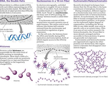
DNA Double Helix: The fundamental structure of DNA, with a diameter of about 2 nm.
Histones: Small, positively charged proteins that bind to DNA and facilitate its compaction. There are five main types: H1, H2A, H2B, H3, and H4.
Nucleosome: About 146 base pairs of DNA wound around a core of eight histone proteins, forming a "bead-on-a-string" structure (10-nm fiber).
Example: Nucleosomes are connected by linker DNA, and histone tails extend outward, playing roles in chromatin structure and gene regulation.
Higher-Order Chromatin Structure
Beyond the nucleosome, chromatin is further compacted through several levels:
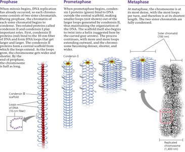
30-nm Fiber: Nucleosomes coil to form a thicker fiber, stabilized by interactions between histone tails and linker DNA.
Looped Domains: The 30-nm fiber forms loops attached to a protein scaffold, further compacting the DNA.
Metaphase Chromosome: During mitosis, chromatin condenses into the highly organized metaphase chromosome, visible under a light microscope.
Chromatin States: Euchromatin and Heterochromatin
Definitions and Functional Differences
Chromatin exists in two main states, which differ in structure and function:
Euchromatin: Loosely packed chromatin that is accessible to the transcription machinery, allowing gene expression. Appears as a diffuse mass in the nucleus during interphase.
Heterochromatin: Densely packed chromatin, often found at centromeres and telomeres. It is generally transcriptionally inactive due to its compact structure.
Table: Comparison of Euchromatin and Heterochromatin
Property | Euchromatin | Heterochromatin |
|---|---|---|
Structure | Loosely packed | Densely packed |
Location | Throughout nucleus | Centromeres, telomeres, specific regions |
Gene Activity | Transcriptionally active | Transcriptionally inactive |
Microscopy Appearance | Diffuse | Irregular clumps |
Example: Genes located in euchromatin are generally expressed, while those in heterochromatin are often silenced.
Dynamic Nature of Chromosomes
Chromatin Remodeling and Cell Cycle
Chromatin structure is dynamic and changes during the cell cycle and in response to cellular needs:
During interphase, chromatin is less condensed, allowing access for DNA replication and transcription.
As the cell prepares for mitosis, chromatin condenses into visible chromosomes to facilitate accurate segregation.
Chemical modifications of histones (e.g., acetylation, methylation) regulate chromatin structure and gene expression.
Additional info: Chromatin remodeling is essential for processes such as DNA repair, replication, and gene regulation. Defects in chromatin structure can lead to diseases, including cancer.
Concept Check
Describe the structure of a nucleosome, the basic unit of DNA packing in eukaryotic cells. A nucleosome consists of a segment of DNA wound around a core of eight histone proteins. This structure compacts DNA and regulates its accessibility.
What two properties, one structural and one functional, distinguish heterochromatin from euchromatin? Structurally, heterochromatin is more densely packed than euchromatin. Functionally, heterochromatin is generally transcriptionally inactive, while euchromatin is transcriptionally active.
