 Back
Chromosome Structure: DNA Packing and Chromatin Organization
Study Guide - Smart Notes
 Tailored notes based on your materials, expanded with key definitions, examples, and context.
Chromosome Structure and DNA Packing
Overview of Chromosome Structure
Chromosomes are highly organized structures composed of DNA and proteins, essential for the storage, expression, and transmission of genetic information. The packaging of DNA into chromosomes allows the long DNA molecules to fit within the cell nucleus and facilitates processes such as gene regulation, replication, and cell division.
DNA in Prokaryotes vs. Eukaryotes
Prokaryotic Chromosomes: Typically consist of a single, circular, double-stranded DNA molecule associated with a small number of proteins. Example: Escherichia coli (E. coli).
Eukaryotic Chromosomes: Composed of single, linear DNA molecules associated with a large number of proteins, forming a complex called chromatin.
Example: The human genome contains DNA molecules that, if stretched out, would measure about 2 meters in length, yet fit within a nucleus only a few micrometers in diameter.
Levels of DNA Packing in Eukaryotic Chromosomes
DNA, the Double Helix
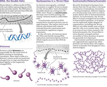
The fundamental structure of DNA is the double helix, with two antiparallel strands wound around each other. This structure is the starting point for higher-order packing.
Histones
Histones are small, positively charged proteins that play a critical role in DNA packaging. There are five main types of histones (H1, H2A, H2B, H3, H4), and they help organize DNA into structural units called nucleosomes.
About one-fifth of a histone's amino acids are positively charged, allowing them to bind tightly to the negatively charged DNA backbone.
Histones are essential for the first level of DNA packing in chromatin.
Nucleosomes and the 10-nm Fiber
The nucleosome is the basic unit of DNA packing in eukaryotes. Each nucleosome consists of DNA wrapped around a core of eight histone proteins (two each of H2A, H2B, H3, and H4). Nucleosomes are connected by short stretches of linker DNA, resembling "beads on a string." This structure forms the 10-nm chromatin fiber.
Nucleosomes help compact DNA and regulate access to genetic information.
Histone tails can be chemically modified, influencing chromatin structure and gene activity.
Higher-Order Chromatin Structure
Chromatin undergoes further folding and coiling to form higher-order structures, especially during cell division:
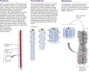
30-nm Fiber: Nucleosomes coil to form a thicker fiber, stabilized by interactions with histone H1 and other proteins.
Looped Domains: The 30-nm fiber forms loops attached to a protein scaffold, further compacting the DNA.
Metaphase Chromosome: During mitosis, chromatin condenses into the highly compact metaphase chromosome, visible under a light microscope.
Chromatin Dynamics and Functional States
Chromatin Changes During the Cell Cycle
Chromatin structure is dynamic and changes throughout the cell cycle:
During interphase, chromatin is less condensed and appears as a diffuse mass in the nucleus, allowing access for gene expression and DNA replication.
As the cell prepares for mitosis, chromatin condenses into distinct chromosomes, facilitating their segregation.
Euchromatin vs. Heterochromatin
Chromatin exists in two main functional states:
Euchromatin: Loosely packed chromatin that is generally accessible for transcription and gene expression.
Heterochromatin: Densely packed chromatin that is transcriptionally inactive and often found at centromeres and telomeres.
Property | Euchromatin | Heterochromatin |
|---|---|---|
Packing Density | Loosely packed | Densely packed |
Gene Activity | Transcriptionally active | Transcriptionally inactive |
Location | Most of the genome | Centromeres, telomeres |
Regulation of Chromatin Structure
Chemical modifications of histones (such as acetylation, methylation, phosphorylation) and DNA (such as methylation) can alter chromatin structure, affecting gene expression and cellular function. These modifications are essential for processes like development, differentiation, and response to environmental signals.
Summary Table: Levels of DNA Packing
Level | Structure | Description |
|---|---|---|
1 | DNA Double Helix | 2-nm diameter; basic genetic material |
2 | Nucleosome (10-nm fiber) | DNA wrapped around histone core |
3 | 30-nm Fiber | Coiling of nucleosomes |
4 | Looped Domains | 30-nm fiber forms loops attached to scaffold |
5 | Metaphase Chromosome | Highly condensed, visible during mitosis |
Concept Check
Describe the structure of a nucleosome, the basic unit of DNA packing in eukaryotic cells. A nucleosome consists of DNA wrapped around a core of eight histone proteins, forming a "bead" on the DNA string. This structure compacts DNA and regulates access to genetic information.
What two properties, one structural and one functional, distinguish heterochromatin from euchromatin? Structurally, heterochromatin is more densely packed than euchromatin. Functionally, heterochromatin is generally transcriptionally inactive, while euchromatin is transcriptionally active.
