 Back
BackCirculation and Gas Exchange in Animals
Study Guide - Smart Notes
 Tailored notes based on your materials, expanded with key definitions, examples, and context.
Circulation and Gas Exchange
Overview of Circulatory and Respiratory Systems
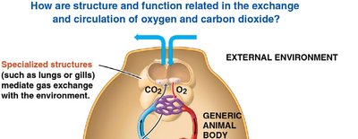
The circulatory and respiratory systems work together to transport oxygen and carbon dioxide between the environment and the cells of the body. Specialized structures such as gills and lungs facilitate gas exchange, while the circulatory system distributes gases and nutrients throughout the organism.
Exchange Surfaces: Structures like gills and lungs increase surface area for efficient gas exchange.
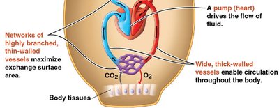
Circulatory System: Consists of a heart (pump), blood vessels, and blood or hemolymph to transport substances.
Diffusion: Small molecules such as O2 and CO2 move between cells and their surroundings by diffusion.
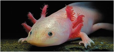
Example: The red feathery appendages on this salamander are gills, specialized for gas exchange in aquatic environments.
Types of Circulatory Systems
Open vs. Closed Circulatory Systems
Animals have evolved two main types of circulatory systems: open and closed. Both systems serve to connect exchange surfaces with body cells, but differ in structure and efficiency.
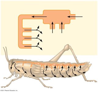
Open Circulatory System: Hemolymph bathes organs directly; found in arthropods and most molluscs.
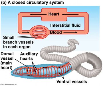
Closed Circulatory System: Blood is confined to vessels; found in annelids, cephalopods, and all vertebrates. More efficient at transporting fluids to tissues and cells.
Basic Components: Circulatory fluid, interconnecting vessels, and a muscular pump (heart).
Vertebrate Circulatory Systems
Single and Double Circulation
Vertebrates display variations in heart structure and circulation patterns, adapted to their metabolic needs.
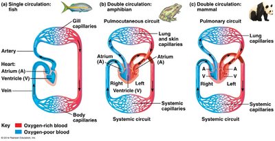
Single Circulation: Found in fish; blood passes through the heart once in each complete circuit.
Double Circulation: Found in amphibians, reptiles, and mammals; blood passes through the heart twice, separating pulmonary (lung) and systemic (body) circuits.
Heart Chambers: Fish have two chambers; amphibians have three; mammals and birds have four.
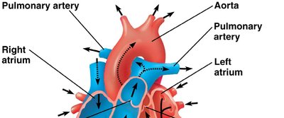
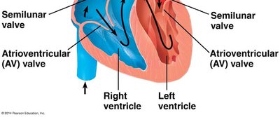
Blood Flow in the Mammalian Heart
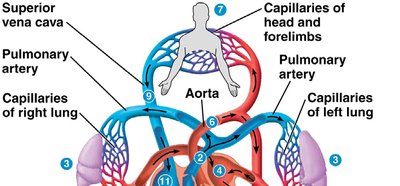
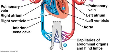
The mammalian heart is a four-chambered organ that separates oxygen-rich and oxygen-poor blood, ensuring efficient delivery of oxygen to tissues.
Right Side: Pumps oxygen-poor blood to the lungs (pulmonary circuit).
Left Side: Pumps oxygen-rich blood to the body (systemic circuit).
Major Vessels: Pulmonary arteries, pulmonary veins, aorta, superior and inferior vena cava.
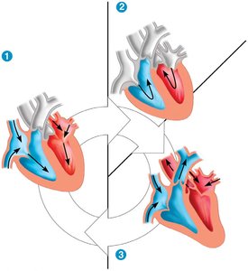
The Cardiac Cycle and Heart Function
Cardiac Cycle
The cardiac cycle consists of alternating periods of contraction (systole) and relaxation (diastole) of the heart chambers. This cycle ensures continuous blood flow throughout the body.
Systole: Contraction phase; blood is pumped out of the chambers.
Diastole: Relaxation phase; chambers fill with blood.
Heart Rate: Number of beats per minute (pulse).
Cardiac Output: Volume of blood pumped by each ventricle per minute.
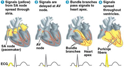
Control of Heart Rhythm
The heart's rhythmic contractions are coordinated by specialized cardiac muscle cells. The sinoatrial (SA) node acts as the pacemaker, initiating electrical impulses that spread through the heart.
SA Node: Sets the pace of the heartbeat.
AV Node: Delays the impulse before passing it to the ventricles.
Purkinje Fibers: Distribute the impulse throughout the ventricles.
Regulation: Nervous system and hormones can influence heart rate.
Blood Vessel Structure and Function
Types of Blood Vessels
Blood vessels are specialized for their roles in circulation. Their structure reflects their function in transporting blood under varying pressures.
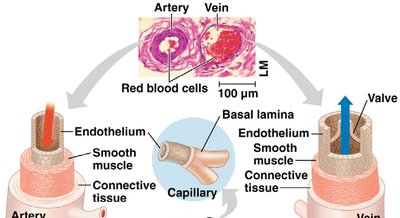
Arteries: Carry blood away from the heart; thick, elastic walls to withstand high pressure.
Veins: Return blood to the heart; thinner walls, valves to prevent backflow.
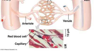
Capillaries: Microscopic vessels where exchange of gases, nutrients, and wastes occurs; thin walls for diffusion.
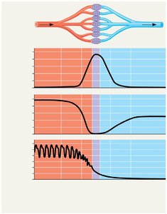
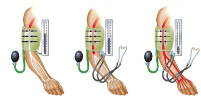
Blood Pressure and Flow
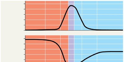
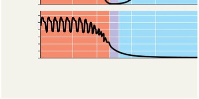
Blood pressure is the force exerted by blood on vessel walls. It is highest in arteries and lowest in veins. Blood flow velocity is slowest in capillaries, allowing time for exchange of materials.
Systolic Pressure: Pressure during ventricular contraction.
Diastolic Pressure: Pressure during ventricular relaxation.
Regulation: Vasoconstriction increases pressure; vasodilation decreases pressure.
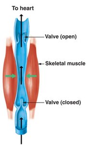
Venous Return and Valves
Blood returns to the heart through veins, aided by skeletal muscle contractions and one-way valves that prevent backflow. Problems with valves can lead to varicose veins.
Capillary Exchange and the Lymphatic System
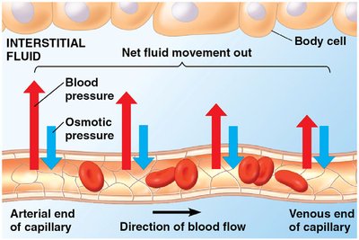
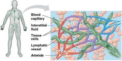
Fluid Exchange in Capillaries
Exchange of substances between blood and interstitial fluid occurs across capillary walls. The balance between blood pressure and osmotic pressure determines the direction of fluid movement.
At Arterial End: Blood pressure exceeds osmotic pressure; fluid moves out.
At Venous End: Osmotic pressure exceeds blood pressure; fluid moves in.

Lymphatic System
The lymphatic system returns excess interstitial fluid to the bloodstream and plays a role in immune defense. Lymph nodes filter lymph and store white blood cells.
Lymph: Fluid collected from tissues and returned to blood.
Lymph Nodes: Filter lymph and house immune cells.
Edema: Swelling caused by disrupted lymph flow.
Blood Composition and Function
Components of Blood
Blood is a connective tissue composed of plasma and cellular elements. Each component has specialized functions in transport, defense, and clotting.
Plasma: Liquid matrix; 90% water, contains ions, proteins (albumin, antibodies, fibrinogen).
Erythrocytes (Red Blood Cells): Transport oxygen using hemoglobin.
Leukocytes (White Blood Cells): Defense and immunity; several types (monocytes, neutrophils, basophils, eosinophils, lymphocytes).
Platelets: Cell fragments involved in blood clotting.
Cardiovascular Diseases
Atherosclerosis, Heart Attack, and Stroke
Cardiovascular diseases are major causes of mortality. Atherosclerosis involves plaque buildup in arteries, leading to reduced blood flow and risk of heart attack or stroke.
Atherosclerosis: Plaque deposits narrow arteries.
Heart Attack: Death of cardiac muscle due to blocked coronary artery.
Stroke: Death of brain tissue due to blocked or ruptured artery in the brain.
Risk Factors: High LDL cholesterol, hypertension, smoking, lack of exercise.
Gas Exchange and Respiratory Surfaces
Mechanisms of Gas Exchange
Gas exchange supplies oxygen for cellular respiration and removes carbon dioxide. It occurs across specialized surfaces such as skin, gills, tracheae, or lungs, depending on the organism.
Partial Pressure: Gases diffuse from regions of higher to lower partial pressure.
Respiratory Surfaces: Adapted for efficient diffusion (thin, moist, large surface area).
Gills and Countercurrent Exchange
Gills are specialized for gas exchange in aquatic animals. Fish gills use a countercurrent exchange system, where blood flows opposite to water, maximizing oxygen uptake.
Lungs and Breathing
Lungs are internal respiratory surfaces found in terrestrial vertebrates. Mammals ventilate lungs by negative pressure breathing, using the diaphragm and rib muscles to change lung volume.
Control of Breathing
Breathing rate is regulated by the brain (medulla oblongata and pons) in response to changes in blood pH, which reflects CO2 levels.
Respiratory Pigments
Hemoglobin and Gas Transport
Respiratory pigments increase the oxygen-carrying capacity of blood. Hemoglobin in vertebrates binds oxygen and helps buffer blood pH. Other animals may use hemocyanin (copper-based pigment).
Bohr Shift: Lower pH (higher CO2) decreases hemoglobin's affinity for O2, facilitating oxygen release in tissues.
CO2 Transport: 7% dissolved in plasma, 23% bound to hemoglobin, 70% as bicarbonate ions.
Summary Table: Comparison of Circulatory Systems
Feature | Open Circulatory System | Closed Circulatory System |
|---|---|---|
Main Fluid | Hemolymph | Blood |
Vessel Structure | Open sinuses | Closed vessels |
Efficiency | Lower | Higher |
Examples | Arthropods, most molluscs | Annelids, cephalopods, vertebrates |
Additional info: This summary integrates and expands upon the provided lecture slides, adding definitions, examples, and academic context for clarity and completeness.
