 Back
BackCirculation and Gas Exchange in Animals
Study Guide - Smart Notes
 Tailored notes based on your materials, expanded with key definitions, examples, and context.
Circulation and Gas Exchange
Overview of Circulatory and Respiratory Systems
The circulatory and respiratory systems work together to transport oxygen and carbon dioxide, as well as nutrients and wastes, throughout the animal body. Specialized structures and coordinated mechanisms ensure efficient exchange and distribution of gases necessary for cellular metabolism.
Circulatory system: Links exchange surfaces (such as lungs or gills) with cells throughout the body.
Respiratory system: Provides specialized surfaces for gas exchange with the environment.
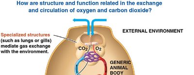
Specialized Exchange Systems
Gills and Simple Body Plans
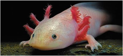
Animals have evolved various adaptations for gas exchange. Gills are specialized structures found in many aquatic animals, such as salamanders, that facilitate the exchange of oxygen and carbon dioxide between the water and blood.
Gills: Outfoldings of the body surface that increase surface area for gas exchange.
Diffusion: Oxygen diffuses from water into blood vessels, while carbon dioxide diffuses from blood into water.
Gastrovascular Cavities in Simple Animals
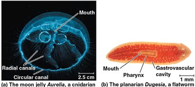
Animals with simple body plans, such as cnidarians and flatworms, use a gastrovascular cavity for both digestion and distribution of substances. This allows direct exchange between the environment and cells by diffusion.
Gastrovascular cavity: Central cavity that functions in both digestion and transport.
Diffusion: Small molecules like O2 and CO2 move between cells and their surroundings.
Types of Circulatory Systems
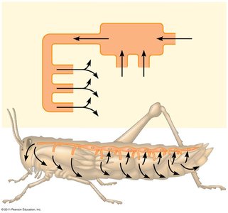
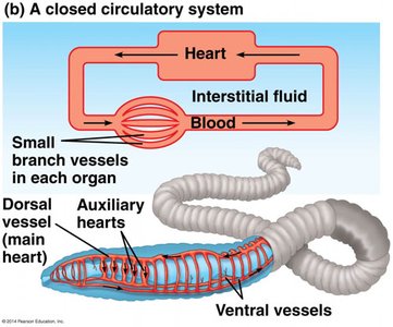
Open and Closed Circulatory Systems
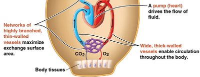
There are two main types of circulatory systems in animals: open and closed. Both systems have three basic components: a circulatory fluid, a set of vessels, and a muscular pump (heart).
Open circulatory system: Hemolymph bathes organs directly; found in insects, other arthropods, and most molluscs.
Closed circulatory system: Blood is confined to vessels and is distinct from interstitial fluid; found in annelids, cephalopods, and all vertebrates.
Vertebrate Circulatory Systems
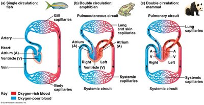
Single and Double Circulation
Vertebrates have evolved different patterns of circulation. Fish have a single circulation system, while amphibians, reptiles, and mammals have double circulation, which separates oxygen-poor and oxygen-rich blood for more efficient oxygen delivery.
Single circulation: Blood passes through the heart once in each complete circuit (e.g., fish).
Double circulation: Blood passes through the heart twice (pulmonary and systemic circuits); found in amphibians, reptiles, and mammals.
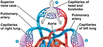
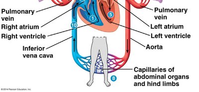
Blood Flow Through the Mammalian Heart
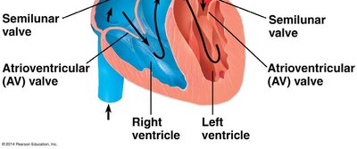
The mammalian heart is a four-chambered organ that ensures separation of oxygen-rich and oxygen-poor blood. The right side pumps blood to the lungs (pulmonary circuit), and the left side pumps blood to the rest of the body (systemic circuit).
Right atrium and ventricle: Receive and pump oxygen-poor blood to the lungs.
Left atrium and ventricle: Receive and pump oxygen-rich blood to the body.
The Cardiac Cycle and Heartbeat Regulation
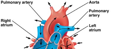
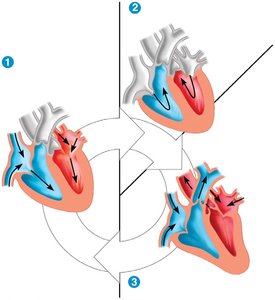
Cardiac Cycle
The cardiac cycle consists of a rhythmic sequence of contraction (systole) and relaxation (diastole) of the heart muscle. This cycle ensures continuous blood flow throughout the body.
Systole: Contraction phase; blood is pumped out of the chambers.
Diastole: Relaxation phase; chambers fill with blood.
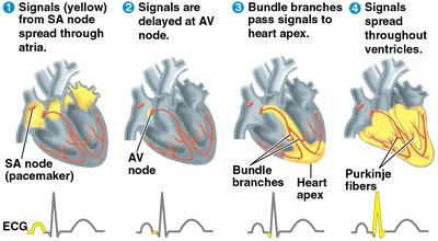
Control of Heart Rhythm
The heart's rhythmic contractions are initiated and coordinated by specialized cardiac muscle cells. The sinoatrial (SA) node acts as the pacemaker, generating electrical impulses that spread through the heart.
SA node: Sets the rate and timing of heart contractions.
AV node: Delays the impulse before passing it to the ventricles.
Purkinje fibers: Distribute the impulse throughout the ventricles.
Blood Vessel Structure and Function
Types of Blood Vessels
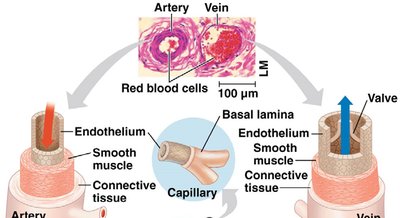
Blood vessels are specialized for their roles in circulation. Arteries, veins, and capillaries differ in structure and function to accommodate blood flow and pressure.
Arteries: Thick-walled vessels that carry blood away from the heart under high pressure.
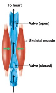
Veins: Thinner-walled vessels that return blood to the heart; contain valves to prevent backflow.
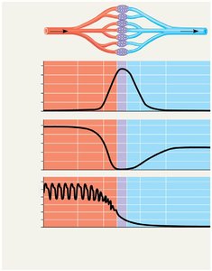
Capillaries: Microscopic vessels with thin walls for exchange of substances with tissues.
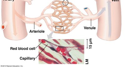
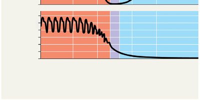
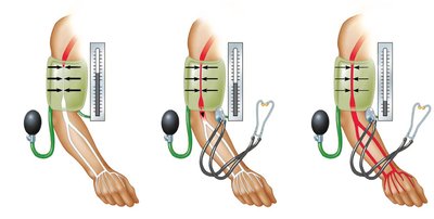
Blood Pressure and Flow
Blood pressure is the force exerted by blood on vessel walls. It is highest in arteries and decreases through arterioles, capillaries, venules, and veins. Blood flow velocity is slowest in capillaries, allowing for exchange of materials.
Systolic pressure: Pressure during ventricular contraction.
Diastolic pressure: Pressure during ventricular relaxation.
Vasoconstriction: Narrowing of blood vessels increases blood pressure.
Vasodilation: Widening of blood vessels decreases blood pressure.
Venous Return and Valves
Blood returns to the heart through veins, aided by skeletal muscle contractions and one-way valves that prevent backflow. Problems with valves can lead to varicose veins.
Capillary Exchange and the Lymphatic System
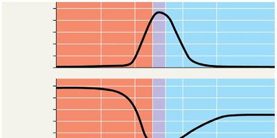
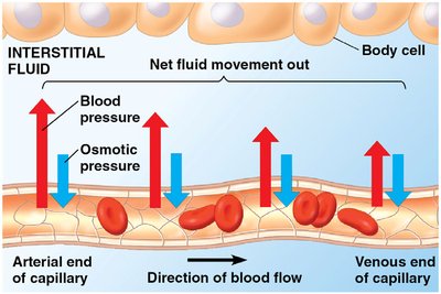
Fluid Exchange in Capillaries
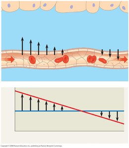
Exchange of substances between blood and interstitial fluid occurs across capillary walls. The balance between blood pressure and osmotic pressure determines the direction of fluid movement.
At the arterial end: Blood pressure exceeds osmotic pressure, causing fluid to leave the capillary.
At the venous end: Osmotic pressure exceeds blood pressure, causing fluid to re-enter the capillary.
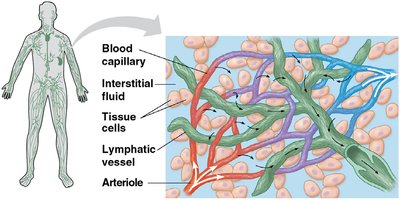
Lymphatic System
The lymphatic system returns excess interstitial fluid to the bloodstream and plays a role in immune defense. Lymph nodes filter lymph and store white blood cells.
Lymph: Fluid collected from tissues and returned to the blood.
Lymph nodes: Filter lymph and house immune cells.
Blood Composition and Function
Components of Blood
Blood is a connective tissue composed of plasma and cellular elements. Plasma is mostly water with dissolved ions and proteins, while cellular elements include erythrocytes, leukocytes, and platelets.
Plasma: 90% water, contains electrolytes, proteins (albumin, antibodies, fibrinogen).
Erythrocytes (red blood cells): Transport oxygen using hemoglobin.
Leukocytes (white blood cells): Defense and immunity.
Platelets: Involved in blood clotting.
Cardiovascular Diseases
Atherosclerosis, Heart Attack, and Stroke
Cardiovascular diseases are disorders of the heart and blood vessels. Atherosclerosis is caused by plaque buildup in arteries, which can lead to heart attacks and strokes.
Heart attack: Death of cardiac muscle due to blocked coronary arteries.
Stroke: Death of brain tissue due to blocked or ruptured arteries in the brain.
LDL ("bad cholesterol"): Promotes plaque formation.
HDL ("good cholesterol"): Reduces cholesterol deposition.
Gas Exchange and Respiratory Surfaces
Mechanisms of Gas Exchange
Gas exchange supplies oxygen for cellular respiration and removes carbon dioxide. Gases diffuse down partial pressure gradients across specialized respiratory surfaces, such as skin, gills, tracheae, or lungs.
Partial pressure: The pressure exerted by a particular gas in a mixture.
Respiratory surfaces: Adapted for efficient gas exchange (e.g., gills in aquatic animals, lungs in terrestrial animals).
Countercurrent Exchange in Fish Gills
Fish gills use a countercurrent exchange system, where blood flows in the opposite direction to water passing over the gills, maximizing oxygen uptake.
Tracheal System in Insects
Insects have a tracheal system, a network of air tubes that deliver oxygen directly to tissues.
Lungs in Mammals
Mammalian lungs are highly branched structures that end in alveoli, where gas exchange occurs. The circulatory system transports gases between the lungs and body tissues.
Breathing and Regulation
Mechanics of Breathing
Mammals ventilate their lungs by negative pressure breathing, using the diaphragm and rib muscles to change lung volume. The medulla oblongata and pons in the brain regulate breathing rate and depth in response to blood pH and CO2 levels.
Respiratory Pigments
Hemoglobin and Gas Transport
Respiratory pigments, such as hemoglobin in vertebrates, bind and transport oxygen. Hemoglobin also helps transport carbon dioxide and buffers blood pH.
Bohr shift: Lower pH decreases hemoglobin's affinity for oxygen, facilitating oxygen release in tissues.
Summary Table: Comparison of Circulatory Systems
System Type | Main Fluid | Vessels | Pump | Examples |
|---|---|---|---|---|
Open | Hemolymph | Open sinuses | Heart (tubular) | Insects, most molluscs |
Closed | Blood | Arteries, veins, capillaries | Heart (chambered) | Annelids, vertebrates |
