 Back
Circulation and Gas Exchange in Animals
Study Guide - Smart Notes
 Tailored notes based on your materials, expanded with key definitions, examples, and context.
Circulation & Gas Exchange
Introduction to Circulation and Gas Exchange
All animals require efficient systems for the exchange of gases (O2 and CO2) to support cellular respiration and ATP production. Gas-exchange organs maximize the rate of O2/CO2 diffusion by providing a large, thin surface area and maintaining a steep partial-pressure gradient. Blood, a specialized tissue, transports gases, nutrients, and wastes throughout the body, with hemoglobin serving as the primary oxygen-carrying protein.
Four Steps of Gas Exchange
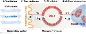
Ventilation: Movement of air or water across a specialized gas-exchange organ.
Gas Exchange: Diffusion of O2 and CO2 between the environment and blood at the respiratory surface.
Circulation: Transport of dissolved gases throughout the body via the circulatory system.
Cellular Respiration: Exchange of gases between blood and tissues, supporting ATP production in mitochondria.
Gas Exchange and Animal Size
Diffusion and Small Animals
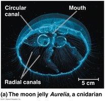
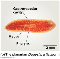
Diffusion time increases with the square of distance, making it efficient only over small distances. Small or thin animals, such as cnidarians and flatworms, lack circulatory or respiratory systems and rely on direct exchange with their environment. Their large surface area to volume ratio and simple body plans facilitate this process.
Circulatory Systems in Large Animals
Larger animals require circulatory systems composed of a circulatory fluid, interconnecting vessels, and a muscular pump (heart). These systems are classified as open or closed, depending on whether the circulatory fluid is confined to vessels.
Open vs. Closed Circulatory Systems
Open Circulatory Systems
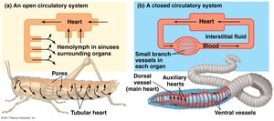
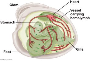
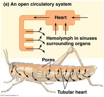
In open circulatory systems, hemolymph is pumped at low pressure and flow rates, not confined exclusively to vessels, and directly bathes tissues. This system is most suitable for sedentary or slow-moving organisms with low oxygen demands, such as many arthropods and most mollusks.
Hemolymph: Circulatory fluid that transports wastes and nutrients, and may contain oxygen-carrying pigments, cells, and clotting agents.
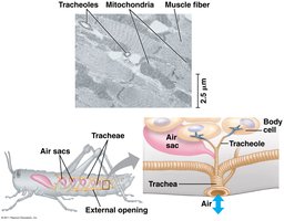
Pancrustaceans: Insects use a tracheal system for direct oxygen delivery, while crustaceans have vessels that can direct hemolymph to tissues with high O2 demand.
Closed Circulatory Systems
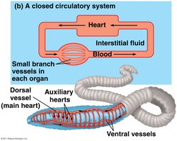
Closed circulatory systems confine blood to vessels, forming a continuous circuit. This allows for high pressure and precise flow, supporting active lifestyles. Closed systems are found in vertebrates, annelids, and cephalopods.
Blood: Circulatory fluid distinct from interstitial fluid, transported through arteries, veins, and capillaries.
Blood Vessels and Circulation
Types of Blood Vessels
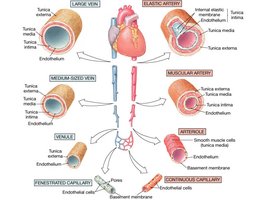
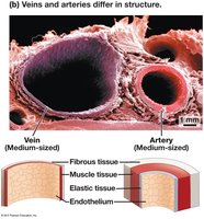
Arteries: Thick-walled vessels carrying blood away from the heart under high pressure.
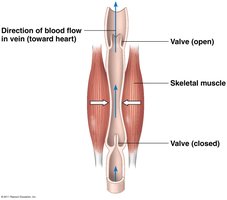
Veins: Thinner-walled vessels returning blood to the heart under lower pressure.
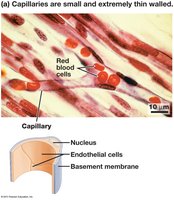
Capillaries: One-cell-thick vessels where exchange of gases, nutrients, and wastes occurs between blood and tissues.
Aorta: The largest artery, originating from the heart.
Arterioles: Small arteries with sphincters regulating blood flow to tissues.
Capillary Exchange and the Lymphatic System
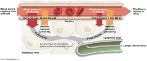
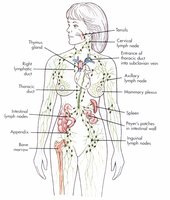
Capillaries are extremely thin and leaky, forming dense networks for exchange. Interstitial fluid builds up due to hydrostatic and osmotic forces. Excess fluid is collected by the lymphatic system and returned to the circulatory system.
Hydrostatic Force: Outward pressure generated by the heart.
Osmotic Force: Inward pressure due to solute concentration differences.
Vertebrate Circulation
Evolution of Heart Structure
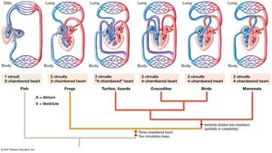
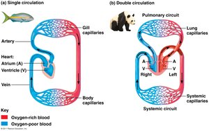
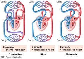
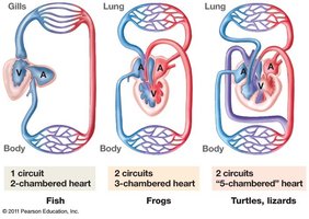
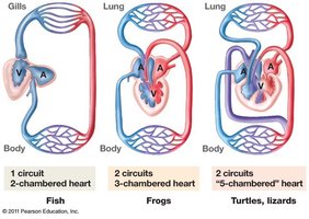
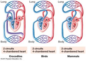
Vertebrate circulatory systems evolved from single-circuit, two-chambered hearts in fish to double-circuit, four-chambered hearts in birds and mammals. This adaptation supports efficient separation of oxygenated and deoxygenated blood, crucial for endothermy and high metabolic rates.
Cardiac Circulation and the Cardiac Cycle
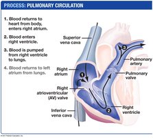
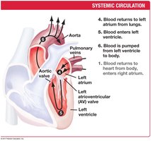
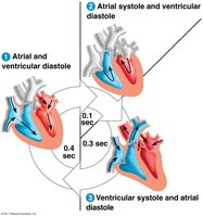
The heart pumps blood through pulmonary (to lungs) and systemic (to body) circuits. The cardiac cycle consists of systole (contraction) and diastole (relaxation), with valves ensuring unidirectional flow. Blood pressure is measured as systolic (peak) and diastolic (resting) values.
Cardiac Output and Heart Regulation
Heart Rate (HR): Beats per minute (~72 bpm in humans).
Stroke Volume (SV): Blood pumped per contraction (~70 ml/beat).
Cardiac Output (CO): Volume of blood pumped per minute (; ~5 L/min in humans).
Pacemaker Cells (SA Node): Initiate heart contraction, regulated by nervous and chemical signals.
AV Node: Relays signal to ventricles for coordinated contraction.
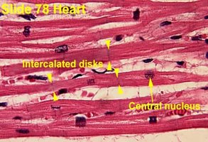
Intercalated Discs: Specialized connections between cardiac muscle cells for rapid electrical signal transmission.
Blood Pressure Regulation
Blood Pressure and Flow
Blood pressure is the force exerted by blood on vessel walls. It drops significantly in capillaries, allowing time for exchange. Baroreceptors in the heart and arteries detect changes in blood pressure, triggering compensatory mechanisms via the nervous system and chemical messengers.
Gas Exchange Mechanisms
Partial Pressure and Diffusion
Gas exchange is driven by differences in partial pressure. Oxygen moves from high partial pressure in the environment to low partial pressure in tissues, while CO2 moves in the opposite direction. The amount of gas dissolved in water depends on solubility, temperature, solute presence, and partial pressure.
Respiratory Structures in Animals
Gills: Outgrowths for gas exchange in aquatic animals, with countercurrent flow maximizing efficiency.
Tracheal System (Insects): Network of tubes delivering air directly to tissues, independent of circulatory system.
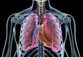
Lungs (Vertebrates): Infoldings of the body surface, with alveoli providing a large surface area for gas exchange.
Ventilation Mechanisms
Amphibians: Use positive pressure to force air into lungs.
Mammals: Use negative pressure via diaphragm contraction for inhalation; exhalation is passive.
Birds: Have unidirectional airflow through lungs, with air sacs and parabronchi maximizing efficiency.
Oxygen Transport and Adaptations
Respiratory Pigments
Hemoglobin: Iron-based pigment in vertebrates and some invertebrates, greatly increasing oxygen-carrying capacity.
Hemocyanin: Copper-based pigment in many arthropods and mollusks.
Adaptations in Diving Mammals
Diving mammals have high blood-to-body volume ratios, store oxygen in myoglobin, and conserve oxygen by reducing muscle blood flow and relying on fermentation during extended dives.
