 Back
BackCirculation and Gas Exchange in Animals
Study Guide - Smart Notes
 Tailored notes based on your materials, expanded with key definitions, examples, and context.
Circulation & Gas Exchange
Overview and Importance
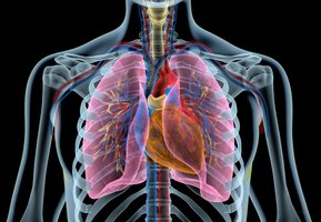
Circulation and gas exchange are essential physiological processes that enable animals to acquire oxygen for cellular respiration and expel carbon dioxide, a metabolic waste product. These processes are tightly linked to the structure and function of respiratory and circulatory systems, which have evolved to maximize the efficiency of gas transport and exchange.
Oxygen is required for ATP production in mitochondria.
Carbon dioxide must be removed to maintain cellular function and pH balance.
Gas-exchange organs maximize diffusion by providing a large, thin surface area and maintaining a steep partial-pressure gradient.
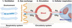
Four Steps of Gas Exchange
Gas exchange in animals involves four key steps, each associated with specific anatomical structures and physiological processes:
Ventilation: Movement of air or water across a specialized gas-exchange surface (e.g., lungs, gills).
Gas Exchange: Diffusion of O2 and CO2 between the environment and blood at the respiratory surface.
Circulation: Transport of dissolved gases throughout the body via the circulatory system.
Cellular Respiration: Exchange of gases between blood and tissues, supporting ATP production in mitochondria.
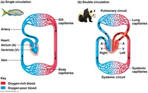
Circulatory Systems: Open vs. Closed
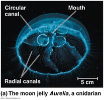
Diffusion and Simple Body Plans
In small or thin animals, such as cnidarians and flatworms, diffusion alone is sufficient for gas and nutrient exchange due to their high surface area to volume ratio and simple body organization.
Cnidarians possess a gastrovascular cavity that functions in both digestion and circulation.
Flatworms have a branched gastrovascular cavity and thin body walls to facilitate exchange.

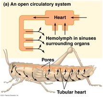
Open Circulatory Systems
Open circulatory systems are characterized by hemolymph (a fluid analogous to blood) that is not entirely confined to vessels. This system is typical of arthropods and most mollusks.
Hemolymph is pumped by the heart into open spaces (sinuses) where it bathes organs directly.
Low pressure and low flow rates are suitable for organisms with low metabolic demands.
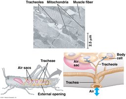
Some pancrustaceans (e.g., insects) have adaptations such as tracheal systems for direct oxygen delivery.
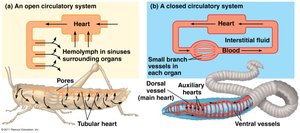
Closed Circulatory Systems
Closed circulatory systems confine blood within vessels, allowing for higher pressure and more efficient, targeted delivery of oxygen and nutrients. This system is found in vertebrates, annelids, and cephalopods.
Blood flows in a continuous circuit of vessels.
High pressure enables rapid and regulated transport.
Supports higher metabolic rates and activity levels.
Blood Vessels and Circulation
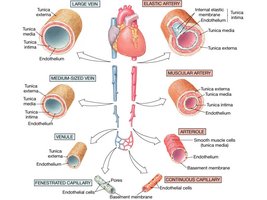
Types of Blood Vessels
Closed circulatory systems contain several types of blood vessels, each with specialized structure and function:
Arteries: Thick-walled vessels that carry blood away from the heart under high pressure.
Veins: Thinner-walled vessels that return blood to the heart under lower pressure.
Capillaries: Extremely thin (one cell layer) vessels where exchange of gases, nutrients, and wastes occurs.
Aorta: The largest artery, originating from the heart.
Arterioles: Small branches of arteries with sphincters to regulate blood flow to tissues.
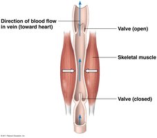
Venous Circulation
Venous return to the heart is facilitated by several mechanisms:
Skeletal muscle contractions compress veins, pushing blood toward the heart.
Internal valves prevent backflow.
Respiratory movements and smooth muscle contractions also assist venous return.
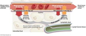
Capillary Exchange and the Lymphatic System
Capillaries allow for the exchange of substances between blood and interstitial fluid. Two main forces govern fluid movement:
Hydrostatic pressure: Outward force generated by blood pressure.
Osmotic pressure: Inward force due to solute concentration in blood plasma.
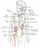
Excess interstitial fluid is collected by the lymphatic system and returned to the circulatory system.
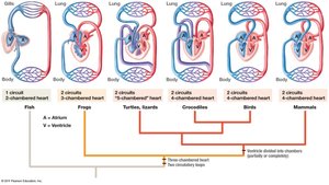
Vertebrate Circulatory System Evolution
Heart Structure and Circuits
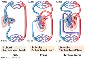
Vertebrate circulatory systems have evolved from single-circuit, two-chambered hearts in fish to double-circuit, four-chambered hearts in birds and mammals. This evolution supports increased metabolic demands and adaptation to terrestrial life.
Fish: Two-chambered heart, single circuit (gills and body).
Amphibians: Three-chambered heart, two circuits (pulmocutaneous and systemic).
Reptiles: Partially divided ventricle, two circuits, some with shunt vessels.
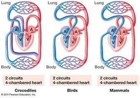
Birds, mammals, crocodiles: Four-chambered heart, complete separation of pulmonary and systemic circuits.
Cardiac Cycle and Blood Flow
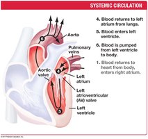
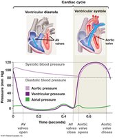
The cardiac cycle consists of alternating periods of contraction (systole) and relaxation (diastole), ensuring unidirectional blood flow through the heart and to the body.
Systole: Ventricular contraction pumps blood into arteries.
Diastole: Heart relaxes and fills with blood.
Valves prevent backflow and produce the characteristic "lub-dup" heart sounds.

Cardiac Output and Regulation
Cardiac output (CO) is the volume of blood pumped by the heart per minute and is determined by heart rate (HR) and stroke volume (SV):
CO = HR × SV
Typical values: HR ≈ 72 beats/min, SV ≈ 70 mL/beat, CO ≈ 5 L/min.
Heart sounds are produced by valve closure; murmurs indicate valve defects.
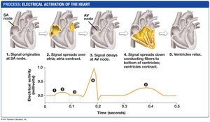
Electrical Control of the Heart
The heart's rhythmic contractions are initiated and coordinated by pacemaker cells in the sinoatrial (SA) node, with signals relayed through the atrioventricular (AV) node and specialized conduction fibers.
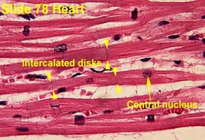
Intercalated discs and gap junctions allow rapid electrical signal transmission between cardiac muscle cells.
Gas Exchange Mechanisms
Principles of Gas Exchange
Gas exchange is driven by differences in partial pressures of oxygen and carbon dioxide between the environment and tissues. Oxygen diffuses from areas of high to low partial pressure, while carbon dioxide diffuses in the opposite direction.
Partial pressure of a gas is calculated as:
Efficient gas exchange requires thin respiratory surfaces and maintenance of steep partial pressure gradients.
Respiratory Adaptations in Animals
Animals have evolved diverse respiratory structures to meet their oxygen demands:
Gills: Outgrowths for aquatic gas exchange, often with countercurrent flow to maximize efficiency.
Tracheal systems: In insects, a network of tubes delivers air directly to tissues.
Lungs: Infolded structures in terrestrial vertebrates, with alveoli providing large surface area for exchange.
Countercurrent Exchange in Fish Gills
Fish gills utilize a countercurrent exchange system, where blood flows opposite to water movement, maintaining a gradient that maximizes oxygen uptake.
Mammalian and Avian Respiration
Mammals use negative pressure breathing, drawing air into lungs via diaphragm contraction. Birds have a highly efficient unidirectional airflow system with air sacs and parabronchi, allowing gas exchange during both inhalation and exhalation.
Respiratory Pigments
Respiratory pigments such as hemoglobin (iron-based) and hemocyanin (copper-based) increase the oxygen-carrying capacity of blood. Hemoglobin is found in vertebrates and some invertebrates, while hemocyanin is present in many arthropods and mollusks.
Adaptations in Diving Mammals
Diving mammals have high blood volume relative to body size, store oxygen in myoglobin, and can reduce oxygen consumption by altering buoyancy and blood flow during dives.
Summary Table: Open vs. Closed Circulatory Systems
Feature | Open Circulatory System | Closed Circulatory System |
|---|---|---|
Main Fluid | Hemolymph | Blood |
Vessel Confinement | Not confined to vessels | Confined to vessels |
Pressure | Low | High |
Organisms | Arthropods, most mollusks | Vertebrates, annelids, cephalopods |
Efficiency | Lower, suitable for low O2 demand | Higher, supports high activity |
