 Back
Circulation and Gas Exchange in Animals
Study Guide - Smart Notes
 Tailored notes based on your materials, expanded with key definitions, examples, and context.
Circulation & Gas Exchange
Overview of Circulatory and Respiratory Systems
The circulatory and respiratory systems work together to facilitate the exchange of gases, nutrients, and waste products between an animal’s cells and its environment. These systems have evolved in complexity across different taxa, with humans possessing a closed circulatory system and highly efficient lungs for gas exchange.
Circulatory system: Transports nutrients, gases, and wastes throughout the body.
Gas exchange: Occurs across specialized surfaces, not to be confused with cellular respiration.
Adaptations: Aquatic and terrestrial organisms have evolved different structures for efficient exchange.
Hemoglobin: A respiratory pigment in red blood cells that increases oxygen-carrying capacity.
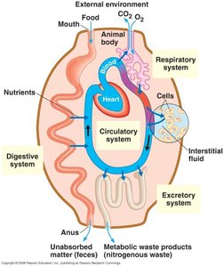
Cellular Exchange with the Environment
Diffusion and the Need for Circulatory Systems
All cells require access to resources and must expel waste. In single-celled and simple multicellular organisms, direct exchange with the environment is possible due to short diffusion distances. In larger, more complex organisms, circulatory systems are necessary to connect isolated cells to the external environment.
Single-celled organisms: All cells are in direct contact with the environment; diffusion is sufficient.
Simple multicellular organisms: All cells are near the body surface or internal cavity, allowing for direct exchange.
Larger multicellular organisms: Circulatory systems transport materials to and from cells.
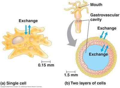
Types of Circulatory Systems
Gastrovascular Cavities and Specialized Circulatory Systems
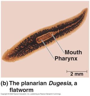
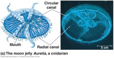
Animals exhibit a range of circulatory system complexities. Some, like cnidarians and flatworms, lack specialized circulatory systems and rely on gastrovascular cavities for exchange. More complex animals have specialized circulatory systems with three main components: circulatory fluid, vessels, and a heart.
Gastrovascular cavity: Highly branched cavity increases surface area for exchange (e.g., Dugesia, Aurelia).
Specialized circulatory system: Consists of circulatory fluid, vessels, and a muscular pump (heart).
Open vs. Closed Circulatory Systems
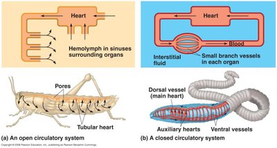
There are two main types of circulatory systems in animals: open and closed. Open systems, found in arthropods and most mollusks, have hemolymph that bathes organs directly. Closed systems, found in annelids and vertebrates, keep blood confined to vessels, allowing for more efficient transport and regulation.
Open circulatory system: Hemolymph is in direct contact with tissues; lower pressure, can serve as a hydrostatic skeleton.
Closed circulatory system: Blood is confined to vessels; allows for faster delivery of oxygen and easier regulation of flow.
Vertebrate Circulatory System Variation
Cardiovascular System Structure
Vertebrates possess a cardiovascular system with a heart (at least two chambers), arteries, veins, and capillaries. The direction of blood flow distinguishes arteries (away from heart) and veins (toward heart).
Heart chambers: Atrium (receives blood), ventricle (pumps blood out).
Arteries and veins: Arteries branch into arterioles and capillaries; veins collect blood from venules.
Single vs. Double Circulation
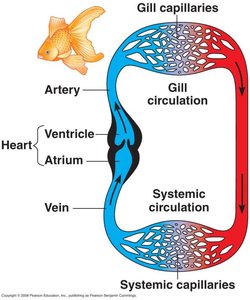
Vertebrates show variation in circulatory circuits. Fish have single circulation with a two-chambered heart, while tetrapods have double circulation with three or four chambers. Double circulation separates pulmonary (lung) and systemic (body) circuits, maintaining higher blood pressure and velocity.
Single circulation: Blood passes through two capillary beds per circuit; lower pressure, aided by muscle movement (e.g., fish).
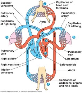
Double circulation: Right side pumps to lungs (pulmonary), left side to body (systemic); higher efficiency (e.g., mammals, birds).
Heart chambers: Amphibians and most reptiles have three chambers; mammals and birds have four.
Pathway of Blood Flow in Mammals
The flow of blood through the mammalian heart and body involves a series of steps that ensure oxygenation and nutrient delivery to tissues, as well as removal of waste products.
Right ventricle pumps blood to lungs via pulmonary arteries.
Blood flows through lung capillaries (gas exchange).
Oxygenated blood returns to left atrium via pulmonary veins.
Left ventricle pumps blood to body via aorta.
Branches supply head, arms, abdomen, and legs.
Deoxygenated blood returns via superior and inferior vena cava to right atrium.
Capillaries and Exchange with Tissues
Capillary Function and Regulation
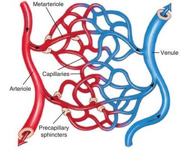
Capillaries are the primary sites of exchange between blood and tissues. Blood flow through capillary beds is regulated to meet tissue needs, controlled by arteriole constriction and precapillary sphincters, under nervous and hormonal control.
Selective flow: Blood is diverted to active tissues (e.g., muscles during exercise, digestive tract after a meal).
Regulation: Achieved by smooth muscle in arterioles and sphincters at capillary entrances.
Mechanisms of Exchange: Pressure and Diffusion
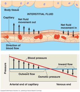
Exchange of gases and solutes occurs by diffusion, while water movement is driven by blood pressure and osmotic pressure. Most water lost from capillaries is recovered; the remainder is returned via the lymphatic system.
Diffusion: O2 and CO2 move down concentration gradients.
Bulk flow: Blood pressure forces water out at arterial end; osmotic pressure draws water in at venous end.
Lymphatic system: Returns excess interstitial fluid to the circulatory system.
The Heart as a Pacemaker
Electrical Control of Heartbeat
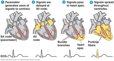
The heart contains autorhythmic cells that generate electrical impulses, coordinating contraction. The sinoatrial (SA) node initiates the heartbeat, which is relayed by the atrioventricular (AV) node after a brief delay, ensuring coordinated contraction of atria and ventricles. The nervous system modulates heart rate according to activity level.
SA node: Initiates contraction (pacemaker).
AV node: Delays signal, then relays to ventricles.
Bundle branches and Purkinje fibers: Distribute signal through ventricles.
Blood Composition
Plasma and Cellular Elements
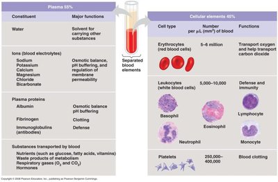
Blood is a connective tissue composed of cells and proteins suspended in plasma. Plasma is mostly water, with dissolved ions, proteins, gases, nutrients, and wastes. Cellular elements include erythrocytes, leukocytes, and platelets.
Plasma: Water, ions (electrolytes), plasma proteins (albumin, fibrinogen, immunoglobulins), nutrients, hormones, wastes.
Cellular elements:
Erythrocytes (red blood cells): Transport oxygen via hemoglobin; lack nuclei and mitochondria.
Leukocytes (white blood cells): Immune defense; several types (e.g., lymphocytes, neutrophils).
Platelets: Cell fragments involved in blood clotting.
Blood Cell Production
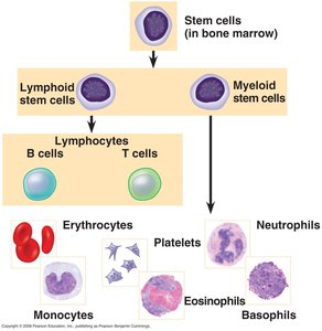
All blood cells are produced from multipotent stem cells in the bone marrow. These stem cells differentiate into various types of blood cells, including erythrocytes, leukocytes, and platelets.
Gas Exchange Across Respiratory Surfaces
Principles of Gas Exchange
Gas exchange occurs by diffusion across moist respiratory surfaces. The rate of diffusion is proportional to surface area and inversely proportional to the thickness of the surface. Oxygen is more available in air than water, but all gas exchange in animals occurs across aqueous surfaces.
Diffusion: Gases move from regions of higher to lower partial pressure.
Surface area: Large, thin surfaces maximize exchange efficiency.
Adaptations in Aquatic and Terrestrial Animals
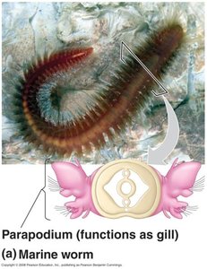
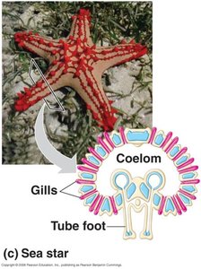
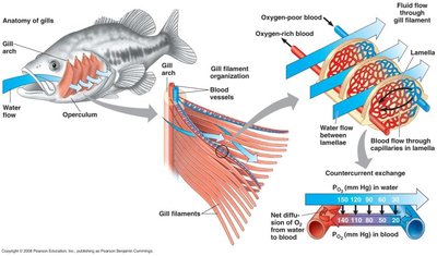
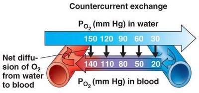
Aquatic animals often have gills—highly vascularized outfoldings of the body surface. Fish gills use countercurrent exchange to maximize oxygen uptake. Terrestrial animals have evolved lungs or tracheal systems for gas exchange.
Gills: Delicate structures with large surface area; countercurrent flow maintains gradient for O2 uptake.
Tracheal system (insects): Network of air tubes delivers O2 directly to tissues.
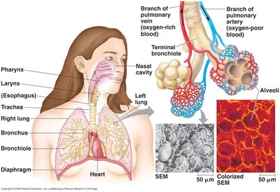
Lungs (vertebrates): Highly branched structures ending in alveoli; site of gas exchange with blood.
Mechanisms of Ventilation
Breathing in Vertebrates
Ventilation is the process of moving respiratory medium (air or water) over the exchange surface. Amphibians use positive pressure breathing, mammals use negative pressure breathing, and birds have a unique system with air sacs for unidirectional flow.
Positive pressure breathing (amphibians): Air is pushed into lungs by oral cavity contraction.
Negative pressure breathing (mammals): Air is drawn into lungs by expanding thoracic cavity using diaphragm and intercostal muscles.
Birds: Use air sacs to maintain continuous, one-way airflow through lungs, maximizing efficiency.
Hemoglobin and Oxygen Transport
Role of Hemoglobin
Oxygen has low solubility in water, so animals use respiratory pigments like hemoglobin to transport O2 efficiently. Hemoglobin, found in erythrocytes, can reversibly bind oxygen, greatly increasing the blood’s oxygen-carrying capacity. The presence of CO2 facilitates the unloading of O2 in tissues.
Structure: Four protein subunits, each with an iron atom that binds O2.
Function: Increases efficiency of oxygen transport, reducing the volume of blood needed to meet metabolic demands.
Additional info: The Bohr effect describes how increased CO2 and lower pH reduce hemoglobin’s affinity for O2, enhancing oxygen delivery to active tissues.
