 Back
Chapter 17: Circulation and Gas Exchange: Structure and Function in Animals
Study Guide - Smart Notes
 Tailored notes based on your materials, expanded with key definitions, examples, and context.
Circulation and Gas Exchange
Overview of Circulatory Systems
The circulatory system links exchange surfaces with all other cells in the body, facilitating the transport of oxygen, carbon dioxide, nutrients, and waste products. Gas exchange occurs primarily by diffusion, but the circulatory system ensures efficient distribution throughout multicellular organisms.
Open Circulatory Systems: Found in arthropods; the circulatory fluid (hemolymph) bathes organs directly, with no distinction between circulatory and interstitial fluids.
Closed Circulatory Systems: Found in annelids, cephalopods, and all vertebrates; blood is confined to vessels and is distinct from interstitial fluid.
Key Functions: Transport of gases, nutrients, hormones, and waste; regulation of body temperature; immune defense.
Vessel Types in Closed Circulatory Systems
Arteries: Large-diameter vessels carrying blood away from the heart.
Arterioles: Smaller branches of arteries leading to capillaries.
Capillaries: Smallest vessels; site of material exchange with interstitial fluid.
Venules: Small vessels collecting blood from capillaries and leading to veins.
Veins: Large-diameter vessels carrying blood towards the heart.
Evolution of the Vertebrate Circulatory System
Vertebrate hearts evolved from a single series of chambers to a double-circuit heart, allowing for separation of oxygen-rich and oxygen-poor blood.
Pulmonary Circuit: Blood flows from the heart to the lungs and back, allowing for gas exchange.
Systemic Circuit: Blood flows from the heart to the rest of the body and back.
Mammalian Heart Structure and Function
The mammalian heart is a four-chambered organ with two atria and two ventricles. Valves ensure unidirectional blood flow:
Atrioventricular (AV) valves: Between atria and ventricles.
Semilunar (SL) valves: Between ventricles and major arteries (aorta/pulmonary arteries).
Blood flow sequence:
Right ventricle pumps O2-poor blood to the lungs.
Blood loads O2 and unloads CO2 in the lungs.
O2-rich blood returns to the left atrium.
Left ventricle pumps blood to the body, where O2 is delivered and CO2 is collected.
O2-poor blood returns to the right atrium.
Cardiac Cycle and Electrical Conduction
The heart's contractions are autorhythmic, meaning they originate within the heart itself, independent of the central nervous system. The sinoatrial (SA) node acts as the pacemaker, setting the timing and rate of contractions. The sequence of electrical conduction is as follows:
Signals from the SA node spread through the atria.
Signals are delayed at the AV node.
Bundle branches pass signals to the heart apex.
Signals spread throughout the ventricles via Purkinje fibers.
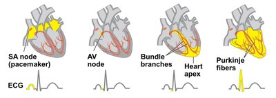
Example: The electrocardiogram (ECG) records the electrical activity of the heart, reflecting the sequence of depolarization and contraction.
Blood Flow Velocity
Blood flow velocity is influenced by vessel diameter. It is slowest in capillaries to facilitate efficient exchange of materials with tissues.
Arteries: Fastest flow due to large diameter and proximity to the heart.
Capillaries: Slowest flow, maximizing exchange time.
Veins: Intermediate flow as blood returns to the heart.
Blood Cell Types and Functions
Red blood cells (erythrocytes): Most numerous; contain hemoglobin for O2 and CO2 transport (some invertebrates use hemocyanin).
White blood cells (leukocytes): Immune response functions; found both inside and outside the circulatory system.
Platelets: Cell fragments that aid in blood clotting (coagulation).
Physiological Respiration
Physiological respiration is the process by which animals obtain oxygen and expel carbon dioxide. Gas exchange occurs across a respiratory surface (layer of epithelial cells) that must remain moist for effective diffusion.
Respiratory medium: The environment where gas exchange occurs (air or water).
Diffusion: Main mechanism for gas movement across respiratory surfaces.
Respiratory Surfaces in Animals
Aquatic animals: Use gills (evaginations) to increase surface area; water must flow constantly for ventilation. Fish use countercurrent flow to maximize exchange efficiency.
Terrestrial animals:
Insects: Tracheal system of branched air tubes; ventilation via abdominal movement; does not require interaction with circulatory system.
Lungs: Invaginations of body surface; amphibians use both lungs and skin; birds have air sacs for continuous airflow; mammals mix fresh and residual air in lungs.
Mammalian Respiratory System
Air enters through the pharynx, passes through the larynx, trachea, bronchi, and bronchioles to reach the alveoli (air sacs at the tips of bronchioles). The respiratory epithelium is lined with cilia and mucus, forming a "mucus escalator" that traps and removes particles.
Gas exchange: Occurs in alveoli; O2 diffuses into capillaries, CO2 diffuses out.
Alveoli: Lack cilia, making them susceptible to contamination.
Mechanisms of Ventilation
Positive pressure breathing: Used by amphibians and birds; air is forced into the lungs.
Negative pressure breathing: Used by mammals; air is drawn into the lungs by expanding the thoracic cavity.
Control of Breathing in Humans
Breathing is regulated by the medulla oblongata in the brain, which uses negative feedback to match metabolic demands. The control is based on blood pH and the O2/CO2 ratio.
Coordination of Circulation and Gas Exchange
Oxygen and carbon dioxide diffuse according to their concentration gradients. High O2 concentration in inhaled air leads to diffusion into the blood, while low CO2 concentration in inhaled air promotes diffusion out of the blood. The process is reversed at the tissue capillaries.
