 Back
Circulation and Gas Exchange: Structure, Function, and Adaptations
Study Guide - Smart Notes
 Tailored notes based on your materials, expanded with key definitions, examples, and context.
Circulatory Systems and Exchange Surfaces
Overview of Circulatory Systems
Circulatory systems are essential for linking exchange surfaces with cells throughout the body, enabling the transport of gases, nutrients, and hormones. As organisms increase in size and complexity, specialized circulatory systems evolve to meet metabolic demands.
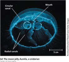
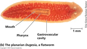
Gastrovascular cavity: A simple body plan where diffusion occurs directly between the environment and cells (e.g., Aurelia, Dugesia).
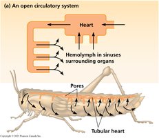
Open circulatory system: Circulatory fluid (hemolymph) bathes organs directly in sinuses; found in arthropods and some mollusks.
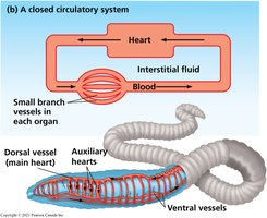
Closed circulatory system: Blood is confined to vessels and is distinct from interstitial fluid; found in annelids, cephalopods, and vertebrates.
Vertebrate Circulatory Systems
Blood Vessel Structure and Function
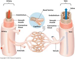
Vertebrate circulatory systems are composed of arteries, veins, and capillaries, each with specialized structures to support their functions.
Arteries: Carry blood away from the heart; thick, elastic walls to withstand high pressure.
Veins: Return blood to the heart; thinner walls and valves to prevent backflow.
Capillaries: Microscopic vessels where exchange of gases, nutrients, and wastes occurs; thin walls for efficient diffusion.
Types of Vertebrate Circulatory Systems
Vertebrates exhibit variations in circulatory system design, reflecting evolutionary adaptations to different metabolic needs.
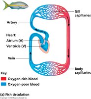
Single circulation: Found in fish; blood passes through the heart once per circuit (heart → gills → body → heart).
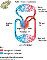
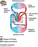
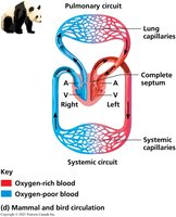
Double circulation: Found in amphibians, reptiles, birds, and mammals; blood passes through the heart twice per circuit, separating pulmonary and systemic circuits.
Heart Structure and Cardiac Cycle
Human Heart Anatomy
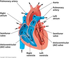
The human heart is a four-chambered organ with two atria and two ventricles. Valves ensure unidirectional blood flow and prevent backflow during contraction.
Atria: Receive blood returning to the heart.
Ventricles: Pump blood out of the heart; thicker muscle walls than atria.
Valves: Atrioventricular (AV) valves and semilunar valves prevent backflow.
Cardiac Cycle and Heart Sounds
The cardiac cycle consists of alternating periods of contraction (systole) and relaxation (diastole). Heart sounds are produced by the closing of valves.
"Lub" sound: Closure of AV valves.
"Dup" sound: Closure of semilunar valves.
Heart murmur: Abnormal sound due to defective valve.
Electrical Conduction and ECG
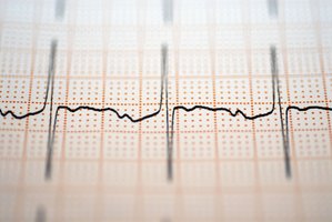
The heart's rhythmic contractions are coordinated by electrical impulses originating from the sinoatrial (SA) node (pacemaker) and transmitted via the atrioventricular (AV) node. The electrocardiogram (ECG) records these impulses.
SA node: Initiates heartbeat and sets pace.
AV node: Delays impulse, allowing atria to contract before ventricles.
ECG: Measures electrical activity of the heart.
Blood Pressure and Blood Flow
Blood Vessel Structure and Flow Dynamics
Blood pressure and flow are influenced by the structure and arrangement of blood vessels. Arteries have thick, elastic walls to accommodate pressure surges, while veins have valves to prevent backflow at low pressure. Capillaries, with their large total surface area, slow blood flow to facilitate exchange.
Measuring Blood Pressure
Blood pressure is measured using a sphygmomanometer. Systolic pressure is recorded when blood first flows through the artery, and diastolic pressure is recorded when blood flows freely.
Term | Definition |
|---|---|
Systolic Pressure | Pressure during ventricular contraction |
Diastolic Pressure | Pressure during ventricular relaxation |
Blood Components and Function
Blood Composition
Blood consists of plasma (55%) and cellular elements (45%). Plasma is mostly water, while cellular elements include erythrocytes, leukocytes, and platelets.
Component | Major Function |
|---|---|
Plasma | Transport of nutrients, hormones, waste; osmotic balance |
Erythrocytes | Oxygen transport via hemoglobin |
Leukocytes | Defense and immunity |
Platelets | Blood clotting |
Blood Clotting Mechanism
When a blood vessel is damaged, platelets adhere to exposed collagen fibers, forming a plug. Clotting factors trigger a cascade that converts fibrinogen to fibrin, forming a stable clot.
Platelets: Initiate clotting by forming a plug.
Fibrinogen: Inactive precursor converted to fibrin.
Fibrin: Forms threads that reinforce the clot.
Gas Exchange and Respiratory Surfaces
Gas Exchange Principles
Gas exchange involves the uptake of O2 and release of CO2 across specialized surfaces. Diffusion is most efficient across large, thin, moist surfaces with short diffusion distances.
Partial pressure: Gases diffuse from regions of higher to lower partial pressure.
Respiratory surfaces: Gills, tracheae, and lungs are adapted for efficient gas exchange.
Adaptations for Gas Exchange
Respiratory pigments such as hemoglobin and myoglobin increase the oxygen-carrying capacity of blood, facilitating efficient gas transport and supporting high metabolic rates.
Hemoglobin: Binds up to four O2 molecules per molecule; found in erythrocytes.
Myoglobin: Stores oxygen in muscle tissue; important in diving mammals.
Summary Table: Circulatory System Types
System Type | Main Fluid | Vessel Structure | Example Organisms |
|---|---|---|---|
Gastrovascular cavity | Body fluid | Single cavity | Jellyfish, flatworms |
Open circulatory system | Hemolymph | Sinuses, tubular heart | Insects, crustaceans |
Closed circulatory system | Blood | Vessels, multi-chambered heart | Annelids, vertebrates |
