 Back
BackCirculation and Gas Exchange: Structure, Function, and Adaptations in Animals
Study Guide - Smart Notes
 Tailored notes based on your materials, expanded with key definitions, examples, and context.
Circulation and Gas Exchange
Overview of Circulatory and Respiratory Systems
The circulatory and respiratory systems are essential for transporting oxygen, nutrients, and removing waste products such as carbon dioxide from animal bodies. These systems have evolved specialized structures and mechanisms to maximize efficiency in gas exchange and internal transport.
Circulatory system: Links exchange surfaces (e.g., lungs, gills) with cells throughout the body.
Respiratory system: Provides specialized surfaces for gas exchange with the environment.
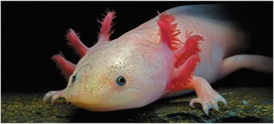
Example: The red feathery appendages on this salamander are gills, specialized for aquatic gas exchange.
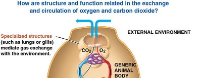
Example: Specialized structures such as lungs or gills mediate gas exchange between the animal and the external environment.
Types of Circulatory Systems
Open vs. Closed Circulatory Systems
Animals have evolved two main types of circulatory systems to distribute materials efficiently:
Open circulatory system: Circulatory fluid (hemolymph) bathes organs directly; found in arthropods and most molluscs.
Closed circulatory system: Blood is confined to vessels and is distinct from interstitial fluid; found in annelids, cephalopods, and all vertebrates.
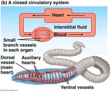
Example: In earthworms, blood circulates within vessels, and auxiliary hearts help pump blood throughout the body.
Vertebrate Circulatory Systems
Single and Double Circulation
Vertebrates display two main patterns of circulation:
Single circulation: Blood passes through the heart once in each complete circuit (e.g., fish).
Double circulation: Blood passes through the heart twice per circuit—once for gas exchange (pulmonary or pulmocutaneous circuit) and once for systemic circulation (e.g., amphibians, reptiles, mammals).
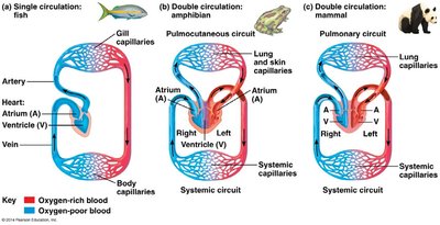
Example: Fish have a two-chambered heart and single circulation, while mammals have a four-chambered heart and double circulation.
Blood Flow Through the Mammalian Heart
The mammalian heart is a four-chambered organ that separates oxygen-rich and oxygen-poor blood, ensuring efficient delivery of oxygen to tissues.
Right side: Pumps oxygen-poor blood to the lungs (pulmonary circuit).
Left side: Pumps oxygen-rich blood to the body (systemic circuit).
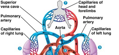
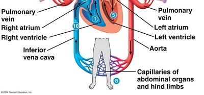
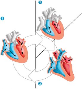
Example: Blood returns from the body via the venae cavae, enters the right atrium, is pumped to the lungs, returns to the left atrium, and is then pumped to the body via the aorta.
Heart Structure and Cardiac Cycle
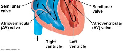
The heart's rhythmic contractions (cardiac cycle) are divided into systole (contraction) and diastole (relaxation). Four valves prevent backflow and ensure unidirectional blood flow.
Atrioventricular (AV) valves: Between atria and ventricles.
Semilunar valves: Between ventricles and major arteries.
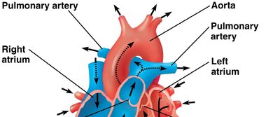
Example: The cardiac cycle consists of atrial and ventricular systole and diastole, coordinating blood flow through the heart.
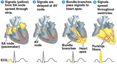
Electrical Control of the Heart
The heart's rhythm is set by the sinoatrial (SA) node (pacemaker), which generates electrical impulses. These impulses spread through the atria, are delayed at the atrioventricular (AV) node, and then travel through specialized fibers to coordinate ventricular contraction.
Blood Vessels: Structure and Function
Types of Blood Vessels
Blood vessels are specialized for their roles in circulation:
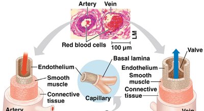
Arteries: Carry blood away from the heart; thick, muscular walls to withstand high pressure.
Veins: Return blood to the heart; thinner walls, valves to prevent backflow.
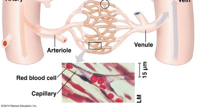
Capillaries: Microscopic vessels for exchange of gases, nutrients, and wastes; thin walls for diffusion.
Blood Pressure and Flow
Blood pressure is the force exerted by blood on vessel walls. It is highest in arteries and lowest in veins. Blood flow velocity is slowest in capillaries, allowing time for exchange.
Regulation of Blood Pressure
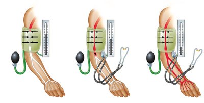
Blood pressure is regulated by cardiac output and peripheral resistance. Vasoconstriction increases pressure, while vasodilation decreases it. Blood pressure can be measured using a sphygmomanometer.
 v
v
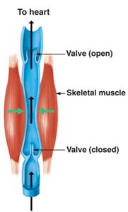
Venous Return and Valves
Blood returns to the heart through veins, aided by skeletal muscle contractions and one-way valves that prevent backflow. Malfunctioning valves can lead to varicose veins.
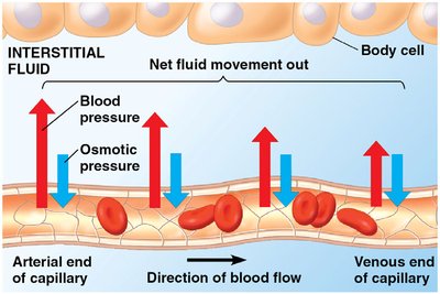
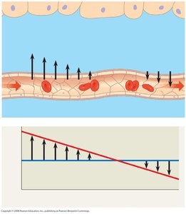
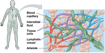
Capillary Exchange and the Lymphatic System
Exchange of substances between blood and interstitial fluid occurs across capillary walls. Fluid movement is driven by the balance of blood pressure and osmotic pressure. The lymphatic system returns excess interstitial fluid to the bloodstream and plays a role in immune defense.
Blood Composition and Function
Components of Blood
Blood is a connective tissue composed of plasma and cellular elements:
Plasma: Liquid matrix (90% water) containing ions, proteins (albumin, antibodies, fibrinogen), nutrients, and wastes.
Cellular elements: Erythrocytes (red blood cells), leukocytes (white blood cells), and platelets (cell fragments for clotting).
Cardiovascular Diseases
Atherosclerosis, Heart Attack, and Stroke
Cardiovascular diseases are major causes of mortality. Atherosclerosis is the buildup of plaques in arteries, which can lead to heart attacks (death of cardiac muscle due to blocked coronary arteries) or strokes (death of brain tissue due to blocked or ruptured arteries).
LDL ("bad cholesterol"): Promotes plaque formation.
HDL ("good cholesterol"): Reduces cholesterol deposition.
Hypertension: High blood pressure increases risk of heart attack and stroke.
Gas Exchange and Respiratory Surfaces
Principles of Gas Exchange
Gas exchange supplies oxygen for cellular respiration and removes carbon dioxide. Gases diffuse down partial pressure gradients across specialized respiratory surfaces, which may include skin, gills, tracheae, or lungs.
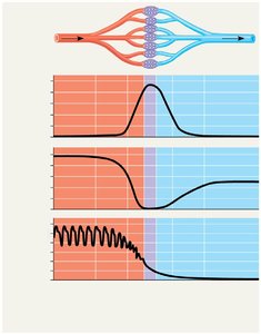
Gills and Countercurrent Exchange
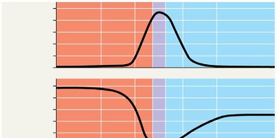
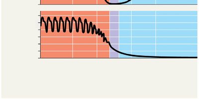
Gills are outfoldings of the body surface specialized for gas exchange in aquatic animals. Fish gills use a countercurrent exchange system, where blood flows opposite to water, maximizing oxygen uptake.
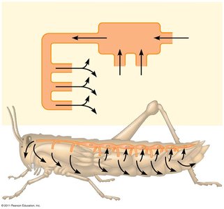
Tracheal Systems in Insects
Insects possess a tracheal system—a network of air tubes that deliver oxygen directly to tissues, independent of the circulatory system.
Lungs and Mammalian Respiration
Lungs are internal, highly vascularized organs for gas exchange in terrestrial vertebrates. Air travels through the respiratory tract to alveoli, where gas exchange occurs with blood in capillaries.
Summary Table: Comparison of Circulatory Systems
System Type | Main Fluid | Vessel Structure | Example Organisms |
|---|---|---|---|
Open | Hemolymph | Sinuses, open spaces | Arthropods, most molluscs |
Closed | Blood | Continuous vessels | Annelids, cephalopods, vertebrates |
Additional info: This guide covers the structure and function of circulatory and respiratory systems, their adaptations, and the physiological principles underlying gas exchange and blood flow in animals.
