 Back
BackCirculation and Gas Exchange: Structure, Function, and Evolution of Cardiovascular and Respiratory Systems
Study Guide - Smart Notes
 Tailored notes based on your materials, expanded with key definitions, examples, and context.
Circulation and Gas Exchange
Overview: The Necessity of Exchange
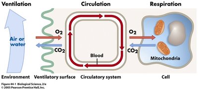
All organisms must exchange materials with their environment to sustain life. These exchanges, including the uptake of oxygen and nutrients and the removal of carbon dioxide and wastes, occur at the cellular level and are mediated by the plasma membrane. In unicellular organisms, exchanges occur directly with the environment, while multicellular organisms rely on specialized systems for efficient transport and exchange.
Mechanisms of Gas Exchange
Diffusion and Convective Transport
Gas exchange relies on diffusion, which is efficient only over small distances and is proportional to the square of the distance traveled. In small or thin animals, cells can exchange materials directly with the environment. Larger or more complex organisms require circulatory systems to transport gases and nutrients efficiently.
Diffusion: Movement of molecules from high to low concentration; rate depends on distance and medium (air or water).
Convective Transport: Bulk movement of fluids (e.g., blood) to enhance exchange rates.
Circulatory Systems: Structure and Function
Open vs. Closed Circulatory Systems
Circulatory systems facilitate the transport of gases, nutrients, and wastes. There are two main types:
Open Circulatory System: Circulating fluid (hemolymph) is not confined to vessels and directly bathes tissues. Example: Tarantulas.
Closed Circulatory System: Blood is confined to vessels, separated from interstitial fluid. Example: Squid.

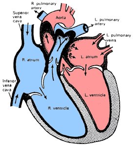
Cardiovascular Structure
The cardiovascular system consists of vessels (arteries, veins, capillaries), blood, and the heart. Vessels are structured with connective tissue, smooth muscle, elastic layers, and an endothelial lining.
Arteries: Carry blood away from the heart; function as pressure reservoirs.
Veins: Return blood to the heart; serve as volume reservoirs.
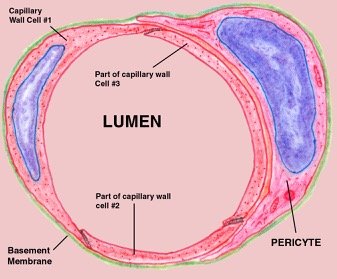
Capillaries: Sites of exchange; walls are a single cell layer thick.


Evolution of Cardiovascular Systems
Single and Double Circulation
Cardiovascular systems have evolved to meet the metabolic demands of different animal groups.
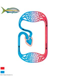
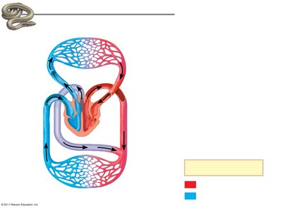
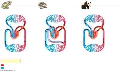
Single Circulation: Found in fish; blood passes through the heart once per circuit (two-chambered heart).
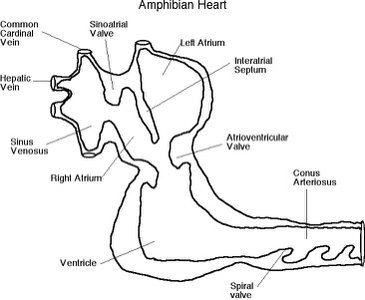
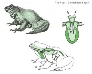
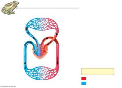
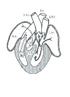
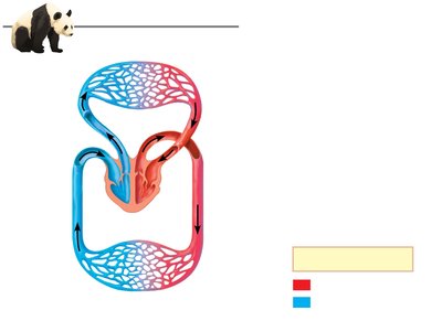
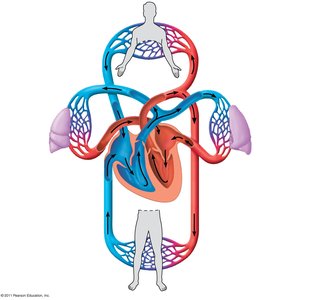
Double Circulation: Found in amphibians, reptiles, birds, and mammals; blood passes through the heart twice per circuit, separating pulmonary and systemic circuits.


Cardiac Cycle and Heart Function
Cardiac Cycle
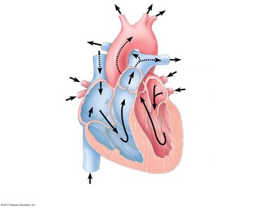
The heart contracts and relaxes in a rhythmic cycle. The contraction phase is called systole, and the relaxation phase is diastole. The heart acts as a dual pump: one side sends deoxygenated blood to the lungs, and the other sends oxygenated blood to the body.
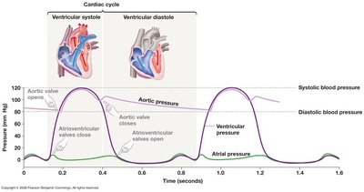
Valves and Cardiac Output
Atrioventricular (AV) Valves: Separate atria and ventricles.
Semilunar Valves: Located at the base of the aorta and pulmonary artery.
Cardiac Output:
Electrical Control of the Heart
Heart contraction is initiated by the sinoatrial (SA) node (pacemaker) and coordinated by the atrioventricular (AV) node, bundle branches, and Purkinje fibers. Electrical activity can be recorded as an electrocardiogram (ECG).
Blood Pressure and Flow
Regulation of Blood Pressure
Systolic Pressure: Pressure during ventricular contraction.
Diastolic Pressure: Pressure during ventricular relaxation.
Vasoconstriction: Increases blood pressure by narrowing arterioles.
Vasodilation: Decreases blood pressure by widening arterioles.
Capillary Exchange and Lymphatic System
Capillary Function
Capillaries are the primary sites of exchange between blood and tissues. Blood flow through capillaries is regulated by precapillary sphincters. Fluid balance is maintained by the lymphatic system, which returns excess fluid to the circulatory system and filters it through lymph nodes.
Blood Composition and Function
Plasma and Cellular Elements
Plasma: Liquid matrix (about 90% water) containing ions, proteins (albumin, fibrinogen, immunoglobulins), nutrients, wastes, gases, and hormones.
Cellular Elements: Erythrocytes (red blood cells), leukocytes (white blood cells), and platelets.
Erythrocytes and Hemoglobin
Erythrocytes: Most numerous blood cells; transport O2 via hemoglobin.
Hemoglobin: Each molecule binds up to four O2 molecules.
Sickle-cell Disease: Caused by abnormal hemoglobin; leads to sickle-shaped cells that can block vessels.
Stem Cells and Blood Cell Production
Stem Cells: Located in red bone marrow; produce erythrocytes, leukocytes, and platelets.
Erythropoietin (EPO): Hormone that stimulates red blood cell production in response to low O2.
Cardiovascular Disease
Atherosclerosis, Heart Attacks, and Stroke
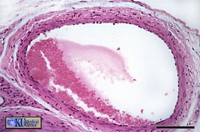
Atherosclerosis: Buildup of plaque in arteries.
Heart Attack (Myocardial Infarction): Death of cardiac muscle due to blocked coronary arteries.
Stroke: Blockage or rupture of arteries in the brain.
Risk Factors: High LDL/HDL ratio, hypertension, diabetes, obesity, smoking, inactivity, age, heredity, stress.
Gas Exchange and Respiratory Systems
Principles of Gas Exchange
Gas exchange supplies O2 for cellular respiration and removes CO2. The efficiency of gas exchange depends on surface area, partial pressure gradients, and the properties of the respiratory medium (air or water).
Partial Pressure: Each gas in a mixture exerts its own pressure; total pressure is the sum of partial pressures (Dalton's Law).
Solubility: O2 is less soluble in water than in air; temperature and solute concentration affect solubility.
Respiratory Organs
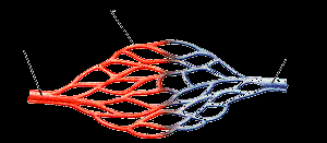
Gills: Outfoldings of the body for aquatic gas exchange; use countercurrent exchange to maximize O2 uptake.
Lungs: Infoldings for terrestrial gas exchange; complexity correlates with metabolic rate.
Mechanisms of Ventilation
Fish: Use buccal pumping or ram ventilation; countercurrent exchange in gills.
Amphibians: Use positive pressure breathing; also exchange gases through skin.
Birds: Have air sacs for unidirectional airflow; cross-current exchange in lungs.
Mammals: Use negative pressure breathing; diaphragm and rib muscles expand thoracic cavity.
Control of Breathing
Medulla Oblongata: Regulates rate and depth of breathing in response to CO2 and pH.
Pons: Regulates breathing tempo.
Negative Feedback: Increased CO2 lowers pH, stimulating increased ventilation.
Respiratory Pigments and Gas Transport
Hemoglobin and Oxygen Transport
Hemoglobin: Increases O2-carrying capacity of blood; each molecule binds four O2 molecules.
Bohr Shift: Lower pH (higher CO2) decreases hemoglobin's affinity for O2, enhancing O2 delivery to tissues.
Carbon Dioxide Transport
About 7% of CO2 is transported dissolved in plasma; most is converted to bicarbonate ions in erythrocytes.
In the lungs, CO2 diffuses out of the blood due to partial pressure gradients.
Adaptations in Diving Mammals
Diving mammals have adaptations such as high blood volume relative to body size, increased myoglobin in muscles, and the ability to tolerate low O2 and high CO2 levels, allowing extended dives.
