 Back
LECTURE 19: Circulatory System and Gas Exchange: Structure, Function, and Adaptation
Study Guide - Smart Notes
 Tailored notes based on your materials, expanded with key definitions, examples, and context.
LECTURE 19
Circulatory System Overview
Major Types of Animal Circulatory Systems
The circulatory system is essential for transporting nutrients, gases, and wastes throughout the body. Animals exhibit a range of circulatory system types, from simple gastrovascular cavities to complex closed systems.
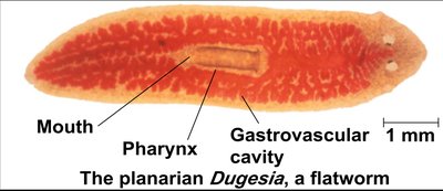
Gastrovascular Cavities: Found in cnidarians and flatworms, these serve both digestive and distribution functions. The body wall is only two cells thick, facilitating diffusion.
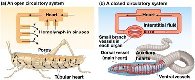
Open Circulatory System: Arthropods and most mollusks have hemolymph that bathes organs directly. Hemolymph is not confined to vessels.
Closed Circulatory System: Annelids, cephalopods, and vertebrates have blood confined to vessels, allowing more efficient transport and regulation.
Vertebrate Circulatory Systems
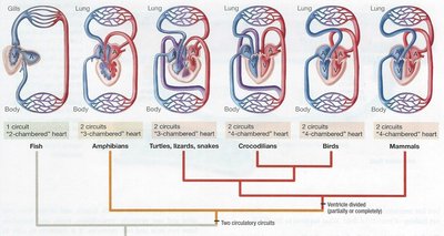
Vertebrates possess a closed cardiovascular system with arteries, veins, and capillaries. The heart structure and circulation patterns vary among groups:
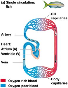
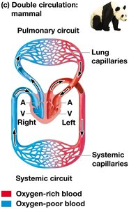
Single Circulation: Seen in fish; blood passes through the heart once per circuit (two-chambered heart).
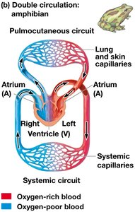
Double Circulation: Found in amphibians, reptiles, birds, and mammals; blood passes through the heart twice per circuit, separating pulmonary and systemic circuits.
Blood: Composition and Function
Blood Components
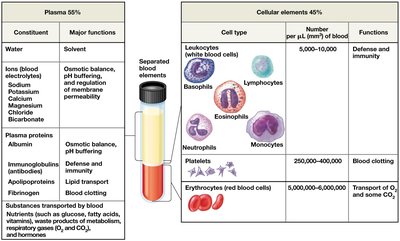
Blood is a connective tissue consisting of cells suspended in plasma. It is responsible for transport, regulation, and defense.
Plasma: The liquid matrix (about 55% of blood) containing water, ions, proteins (albumin, immunoglobulins, fibrinogen), nutrients, hormones, and waste products.
Cellular Elements: About 45% of blood volume, including erythrocytes (red blood cells), leukocytes (white blood cells), and platelets.
Red Blood Cells (Erythrocytes)
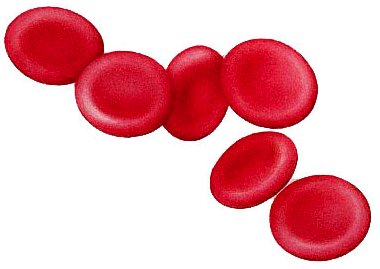
Red blood cells are the most numerous blood cells and are specialized for oxygen transport via hemoglobin. In mammals, they lack nuclei and mitochondria.
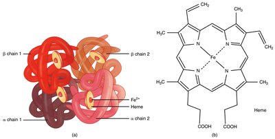
Hemoglobin: An iron-containing protein that binds up to four O2 molecules per molecule.
White Blood Cells (Leukocytes) and Platelets
Leukocytes are involved in defense and immunity. There are five major types: monocytes, neutrophils, basophils, eosinophils, and lymphocytes. Platelets are cell fragments essential for blood clotting.
Leukocytes: Function in phagocytosis and immune responses.
Platelets: Involved in the formation of blood clots.
Blood Cell Formation (Hematopoiesis)
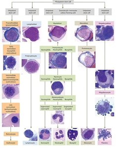
All blood cells develop from multipotent stem cells in the red bone marrow. Erythropoietin (EPO) stimulates red blood cell production in response to low oxygen levels.
Blood Clotting
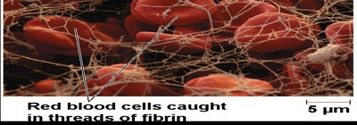
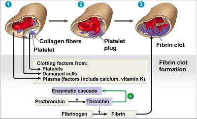
Blood clotting (coagulation) is a cascade of reactions that converts fibrinogen to fibrin, forming a mesh that traps blood cells and seals wounds.
Platelet Plug Formation: Platelets adhere to damaged tissue and aggregate.
Fibrin Clot Formation: Thrombin converts fibrinogen to fibrin, stabilizing the clot.
Blood Types and Compatibility
Blood Types
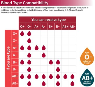
Blood types are determined by the presence or absence of specific antigens on the surface of red blood cells. The ABO and Rh systems are the most clinically significant.
ABO System: Four main types: A, B, AB, O.
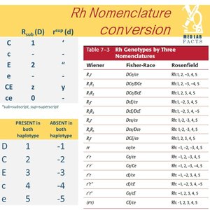
Rh Factor: Presence (+) or absence (−) of the D antigen.
Blood Vessel Structure and Function
Types of Blood Vessels
Blood vessels are classified based on their structure and function:
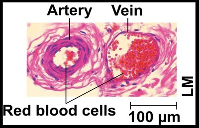
Arteries: Carry blood away from the heart; thick, elastic walls.
Veins: Return blood to the heart; thinner walls, valves prevent backflow.
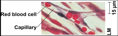
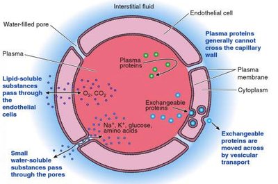
Capillaries: Microscopic vessels where exchange of substances occurs between blood and tissues.
Cardiac Cycle and Heart Function
Heart Structure and Blood Flow
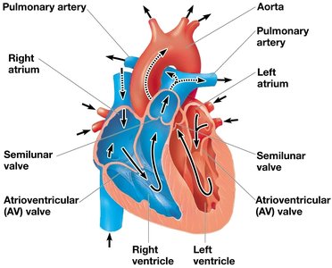
The mammalian heart has four chambers: two atria and two ventricles. Blood flows in one direction, regulated by valves.
Atria: Receive blood returning to the heart.
Ventricles: Pump blood out of the heart.
Cardiac Cycle
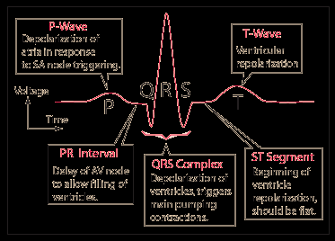
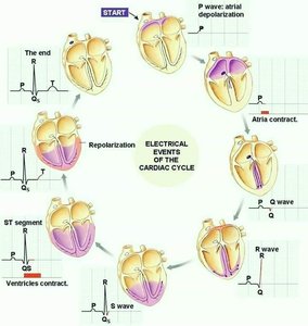
The cardiac cycle consists of systole (contraction) and diastole (relaxation). The sinoatrial (SA) node acts as the pacemaker, initiating electrical impulses that coordinate contraction.
Electrocardiogram (ECG/EKG): Records the electrical activity of the heart.
Gas Exchange and Respiratory Adaptations
Respiratory Surfaces and Media
Gas exchange occurs across specialized, moist respiratory surfaces. Animals may use air or water as the respiratory medium. Efficiency is crucial, especially in water where O2 is less available.
Gills, Tracheae, and Lungs
Gills: Outfoldings of the body surface; fish use countercurrent exchange to maximize O2 uptake.
Tracheal System: In insects, a network of tubes delivers O2 directly to cells.
Lungs: Infoldings of the body surface; complexity correlates with metabolic rate.
Respiratory Pigments
Hemoglobin and Oxygen Transport
Respiratory pigments increase the oxygen-carrying capacity of blood. Hemoglobin (in vertebrates) binds O2 at iron-containing heme groups. Hemocyanin (in some invertebrates) uses copper.
Bohr Shift: Lower pH and higher CO2 decrease hemoglobin's affinity for O2.
CO2 Transport: Most CO2 is transported as bicarbonate ions in plasma.
Hemoglobinopathies
Genetic Disorders of Hemoglobin
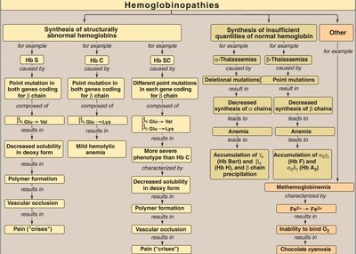
Hemoglobinopathies are inherited disorders affecting the structure or production of hemoglobin, leading to diseases such as sickle cell anemia and thalassemias.
Sickle Cell Disease: Caused by a point mutation in the β-globin gene, resulting in abnormal hemoglobin (HbS) and sickled red cells.
Thalassemias: Caused by mutations leading to decreased synthesis of α or β chains, resulting in anemia.
Methemoglobinemia: Characterized by the presence of oxidized hemoglobin (Fe3+), reducing oxygen delivery.
Summary Table: Blood Components
Component | Major Functions |
|---|---|
Plasma (55%) | Solvent, osmotic balance, pH buffering, transport of nutrients, hormones, and waste |
Red Blood Cells (Erythrocytes) | Transport of O2 and some CO2 |
White Blood Cells (Leukocytes) | Defense and immunity |
Platelets | Blood clotting |
Additional info: This table summarizes the main components and functions of blood as described in the lecture and images.
