 Back
lecture 2 Circulatory Systems in Animals: Structure, Function, and Regulation
Study Guide - Smart Notes
 Tailored notes based on your materials, expanded with key definitions, examples, and context.
lecture 2 Circulatory Systems in Animals
Introduction to Circulatory Systems
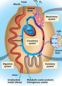
The circulatory system is essential for transporting nutrients, gases, and wastes throughout the bodies of animals. It connects the external environment with internal tissues, ensuring that all cells receive the materials they need for survival and proper function.
Unicellular organisms rely on direct diffusion for exchange of materials with their environment.
Multicellular organisms require specialized circulatory systems to overcome the limitations of diffusion over large distances.
The time for diffusion increases with the square of the distance: (where t is time, x is distance).
Types of Circulatory Systems
Gastrovascular Cavities
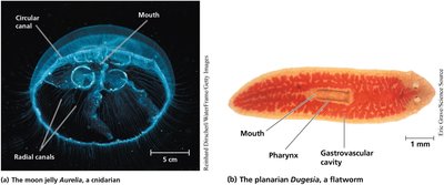
Some simple animals, such as cnidarians and flatworms, use a gastrovascular cavity for both digestion and internal transport. This system minimizes diffusion distances and is adequate for organisms with thin body walls.
Gastrovascular cavity: A central cavity with a single opening that functions in both digestion and distribution of substances.
Examples: Aurelia (moon jelly), Dugesia (planarian flatworm).
Open Circulatory Systems
In open circulatory systems, the circulatory fluid (hemolymph) is not entirely contained within vessels. Instead, it bathes the organs directly in body cavities (sinuses).
Hemolymph: The combined fluid of blood and interstitial fluid.
Found in arthropods (e.g., insects) and most molluscs.
Pressure dissipates quickly, requiring faster heart rates or accessory hearts.

Closed Circulatory Systems
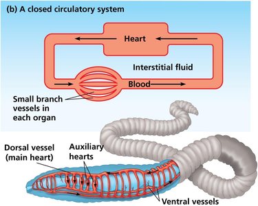
Closed circulatory systems confine blood to vessels, separating it from interstitial fluid. This allows for more efficient transport and higher pressure gradients.
Found in annelids, cephalopods, and all vertebrates.
Blood is pumped by the heart through a continuous circuit of vessels.
Allows for better regulation of blood flow to different tissues.
Organization of Vertebrate Circulatory Systems
Single Circulation
Single circulation is found in fishes and some other aquatic vertebrates. Blood passes through the heart once in each complete circuit.
Two-chambered heart: one atrium, one ventricle.
Blood flows from the heart to the gills (for oxygenation), then to the rest of the body, and back to the heart.
Lower pressure and slower delivery of oxygen to tissues.
Double Circulation: Amphibians
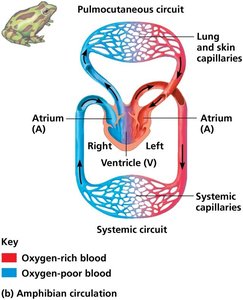
Amphibians have a three-chambered heart and a double circulation system, which separates oxygen-poor and oxygen-rich blood to some extent.
Three chambers: two atria, one ventricle.
Blood is pumped into two circuits: the pulmocutaneous circuit (lungs and skin) and the systemic circuit (body).
Some mixing of oxygenated and deoxygenated blood occurs in the ventricle.
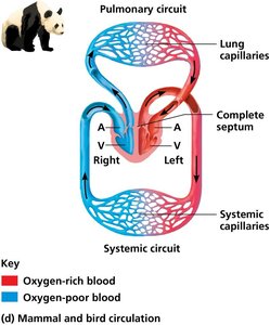
Double Circulation: Mammals and Birds
Mammals and birds have a four-chambered heart, which completely separates oxygen-rich and oxygen-poor blood, supporting high metabolic rates.
Four chambers: two atria, two ventricles.
Two separate circuits: pulmonary (lungs) and systemic (body).
Efficient oxygen delivery supports endothermy (warm-bloodedness).
Human Cardiovascular System
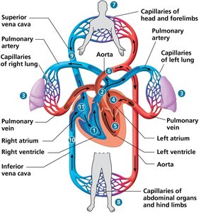
The human cardiovascular system is a closed, double circulatory system with a four-chambered heart. It includes major arteries, veins, and capillaries that ensure efficient transport of blood throughout the body.
Pulmonary circuit: Right ventricle pumps deoxygenated blood to the lungs; oxygenated blood returns to the left atrium.
Systemic circuit: Left ventricle pumps oxygenated blood to the body; deoxygenated blood returns to the right atrium.
Arteries carry blood away from the heart; veins return blood to the heart.
Structure and Function of the Heart
Heart Chambers and Valves
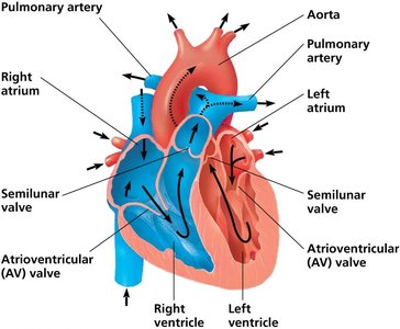
The heart is composed of two atria and two ventricles. Valves ensure unidirectional blood flow and prevent backflow.
Atria: Thin-walled chambers that receive blood returning to the heart.
Ventricles: Thick-walled chambers that pump blood out of the heart.
Atrioventricular (AV) valves: Separate atria from ventricles.
Semilunar valves: Separate ventricles from major arteries.
The Cardiac Cycle
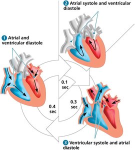
The cardiac cycle consists of alternating periods of contraction (systole) and relaxation (diastole) that pump blood through the heart and into the circulatory system.
Systole: Ventricles contract, pumping blood out of the heart.
Diastole: Heart muscle relaxes, allowing chambers to fill with blood.
Valves open and close in response to pressure changes.
Coordination and Regulation of the Heartbeat
Electrical Conduction System
The heart's rhythmic contractions are coordinated by specialized cardiac muscle cells that generate and conduct electrical impulses.
Sinoatrial (SA) node: The pacemaker of the heart, initiates each heartbeat.
Atrioventricular (AV) node: Delays the impulse before passing it to the ventricles.
Bundle branches and Purkinje fibers: Distribute the impulse throughout the ventricles.
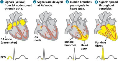
Electrocardiogram (ECG)
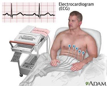
An electrocardiogram (ECG) records the electrical activity of the heart and is used to diagnose heart conditions.
Detects the timing and strength of electrical signals during the cardiac cycle.
Commonly used in clinical settings to monitor heart health.
Blood Vessels and Blood Flow
Structure of Blood Vessels
Blood vessels are specialized for their functions in transporting blood throughout the body.
Arteries: Thick-walled vessels that carry blood away from the heart under high pressure.
Veins: Thinner-walled vessels that return blood to the heart; contain valves to prevent backflow.
Capillaries: Microscopic vessels with thin walls for exchange of materials between blood and tissues.
Blood Pressure and Regulation
Blood pressure is the force exerted by blood on vessel walls. It is highest in arteries and decreases through the circulatory system.
Systolic pressure: Pressure during ventricular contraction.
Diastolic pressure: Pressure during ventricular relaxation.
Regulated by vessel diameter (vasoconstriction and vasodilation), hormones, and nervous system input.
Blood Components and Function
Plasma and Cellular Elements
Blood is a connective tissue composed of plasma and cellular elements.
Plasma: The liquid matrix, mostly water, containing proteins, ions, and nutrients.
Erythrocytes (red blood cells): Transport oxygen using hemoglobin.
Leukocytes (white blood cells): Defend against infection and disease.
Platelets: Cell fragments involved in blood clotting.
Blood Clotting
Blood clotting (coagulation) prevents excessive blood loss after injury.
Platelets adhere to damaged vessels and form a plug.
A cascade of reactions converts fibrinogen to fibrin, forming a stable clot.
Improper clotting can lead to thrombus formation and blockages.
Cardiovascular Disease
Atherosclerosis, Heart Attacks, and Stroke
Cardiovascular diseases are a leading cause of death and are often related to the buildup of fatty deposits in arteries (atherosclerosis).
Atherosclerosis: Plaque buildup narrows arteries, reducing blood flow.
Heart attack (myocardial infarction): Blockage of coronary arteries damages heart muscle.
Stroke: Blockage or rupture of arteries in the brain causes tissue death.
Risk factors include high LDL cholesterol, inflammation, and hypertension.
The Lymphatic System
Role in Circulation and Immunity
The lymphatic system returns excess interstitial fluid to the bloodstream and plays a key role in immune defense.
Lymph vessels collect and return fluid to veins in the neck.
Lymph nodes filter lymph and house immune cells.
Edema results from disruptions in lymph flow.
