Back
Comprehensive Study Guide: Cell Structure, Function, and Microscopy
Study Guide - Smart Notes
 Tailored notes based on your materials, expanded with key definitions, examples, and context.
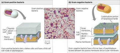
Q1. Describe an overview of key cell structures that are necessary to perform life functions.
Background
Topic: Cell Structure and Function
This question is testing your understanding of the essential components found in all cells and their roles in maintaining life processes.
Key Terms:
Plasma Membrane: Regulates what enters and exits the cell.
Cytoplasm: Gel-like substance where cellular processes occur.
DNA: Genetic material that directs cell activities.
Ribosomes: Sites of protein synthesis.
Step-by-Step Guidance
List the structures found in all cells (plasma membrane, cytoplasm, DNA, ribosomes).
Briefly describe the function of each structure.
Explain why each structure is essential for cell survival and function.
Try solving on your own before revealing the answer!
Q2. Using the table to the right, give the specific cell size ranges for the following and rank them smallest to largest: atom, ribosome, small bacteria, mitochondria, animal cells, chicken egg.
Background
Topic: Cell Size and Scale
This question tests your ability to compare the sizes of biological structures and understand their relative scale.
Key Terms:
Nanometer (nm): 1 billionth of a meter.
Micrometer (μm): 1 millionth of a meter.
Step-by-Step Guidance
Identify the approximate size range for each structure (refer to textbook or class notes if needed).
Convert all sizes to the same unit (e.g., nanometers or micrometers) for easy comparison.
Rank the structures from smallest to largest based on their size.
Try solving on your own before revealing the answer!
Q3. Describe the difference between the different microscopes listed below. Give examples of the structures that are visible by each type of microscope and the rationale on why they were used.
Background
Topic: Microscopy
This question is about understanding the capabilities and uses of different types of microscopes in cell biology.
Key Terms and Types:
Light Microscope: Uses visible light to view cells and large organelles.
Fluorescence Microscope: Uses fluorescent dyes to label and visualize specific cell components.
Electron Microscope: Uses electron beams for high-resolution images of small structures.
Scanning Electron Microscope (SEM): Shows surface details.
Transmission Electron Microscope (TEM): Shows internal structures.
Step-by-Step Guidance
Define each type of microscope and its principle of operation.
List examples of cell structures visible with each microscope (e.g., light microscope for cells, electron microscope for ribosomes).
Explain why a particular microscope is chosen for a specific observation (resolution, sample preparation, etc.).
Try solving on your own before revealing the answer!
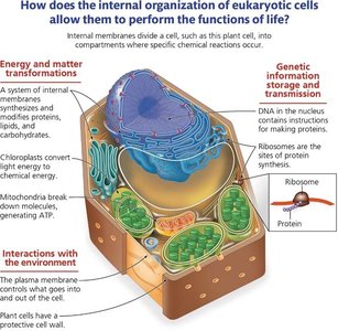
Q8. Why must cells remain small? Explain using the surface area-to-volume ratio.
Background
Topic: Cell Size and Efficiency
This question tests your understanding of how cell size affects the efficiency of material exchange and cellular processes.
Key Terms and Formulas:
Surface Area: The total area of the cell's membrane.
Volume: The space inside the cell.
Surface Area-to-Volume Ratio:
Step-by-Step Guidance
Recall that as a cell grows, its volume increases faster than its surface area.
Explain why a high surface area-to-volume ratio is important for efficient exchange of materials (nutrients, waste, gases).
Describe what happens to this ratio as the cell gets larger and why this limits cell size.
Try solving on your own before revealing the answer!
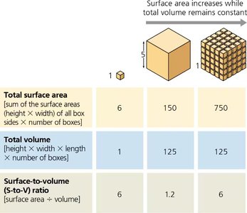
Q17. Using the figure, describe the structural differences in gram-positive vs gram-negative bacteria.
Background
Topic: Bacterial Cell Walls
This question is about distinguishing between two major types of bacterial cell walls and understanding their implications for antibiotic treatment.
Key Terms:
Gram-Positive Bacteria: Thick peptidoglycan layer, no outer membrane.
Gram-Negative Bacteria: Thin peptidoglycan layer, outer membrane present.
Step-by-Step Guidance
Identify the main structural components of each type (peptidoglycan thickness, presence of outer membrane).
Explain how these differences affect staining and susceptibility to antibiotics like penicillin.
Relate the structure to the function and protection of the bacterial cell.
Try solving on your own before revealing the answer!
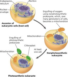
Q25. Describe the endosymbiosis theory and how the current eukaryotic cell may have derived with early origins with a prokaryotic cell.
Background
Topic: Evolution of Eukaryotic Cells
This question tests your understanding of the endosymbiosis theory, which explains the origin of mitochondria and chloroplasts in eukaryotic cells.
Key Terms:
Endosymbiosis: A symbiotic relationship where one organism lives inside another.
Mitochondria and Chloroplasts: Organelles thought to have originated from free-living prokaryotes.
Step-by-Step Guidance
Describe the process by which a host cell engulfed a prokaryotic cell.
Explain how this led to the development of mitochondria (and later, chloroplasts in plant cells).
Discuss the evidence supporting this theory (e.g., double membranes, own DNA, similarity to bacteria).
Try solving on your own before revealing the answer!
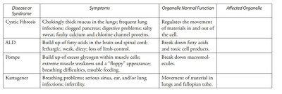
Q50. What organelles or cell structures are likely malfunctioning in these diseases?
Background
Topic: Organelle Function and Disease
This question asks you to connect symptoms of diseases to the malfunction of specific organelles or cell structures.
Key Terms:
Cystic Fibrosis: Involves ion channel proteins in the plasma membrane.
ALD: Involves peroxisomes (breakdown of fatty acids).
Pompé Disease: Involves lysosomes (breakdown of glycogen).
Kartagener Syndrome: Involves cilia (microtubules).
Step-by-Step Guidance
Read the symptoms and match them to the normal function of organelles.
Identify which organelle's malfunction would cause the described symptoms.
Explain how the malfunction leads to the disease symptoms.
Try solving on your own before revealing the answer!