 Back
BackCoordinated Cycles of Heart Contraction and Double Circulation in Mammals
Study Guide - Smart Notes
 Tailored notes based on your materials, expanded with key definitions, examples, and context.
Coordinated Cycles of Heart Contraction Drive Double Circulation in Mammals
Overview of Mammalian Circulation
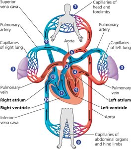
The mammalian cardiovascular system is organized into two main circuits: the pulmonary circuit and the systemic circuit. These circuits operate simultaneously, ensuring efficient oxygenation of blood and delivery of nutrients throughout the body.
Pulmonary Circuit: Carries deoxygenated blood from the right ventricle to the lungs via the pulmonary arteries. In the lungs, blood releases carbon dioxide and picks up oxygen.
Systemic Circuit: Transports oxygenated blood from the left ventricle through the aorta to the rest of the body, delivering oxygen and nutrients to tissues and collecting waste products.
The two ventricles contract almost simultaneously, pumping equal volumes of blood through their respective circuits.
The systemic circuit contains a greater total volume of blood compared to the pulmonary circuit due to its extensive network of vessels.
The Human Heart: Structure and Function
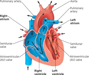
The human heart is a muscular organ located behind the sternum. It consists of four chambers: two atria (upper chambers) and two ventricles (lower chambers). The heart's structure ensures unidirectional blood flow and efficient separation of oxygenated and deoxygenated blood.
Atria: Thin-walled chambers that receive blood returning to the heart from the lungs (left atrium) or the body (right atrium).
Ventricles: Thick-walled chambers that pump blood out of the heart. The left ventricle has the thickest wall to generate the force needed for systemic circulation.
Valves: Four main valves prevent backflow of blood: atrioventricular (AV) valves between atria and ventricles, and semilunar valves at the exits of the ventricles (pulmonary and aortic valves).
The Cardiac Cycle
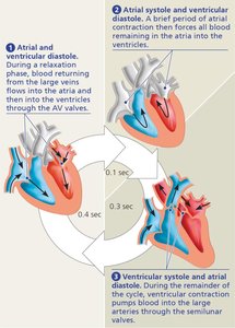
The cardiac cycle refers to the sequence of events during one complete heartbeat, including periods of contraction (systole) and relaxation (diastole) of the heart chambers.
Systole: The contraction phase, during which blood is pumped out of the chambers.
Diastole: The relaxation phase, during which the chambers fill with blood.
One cardiac cycle in a resting adult human lasts about 0.8 seconds.
Cardiac Output: The volume of blood each ventricle pumps per minute, calculated as:
Heart Rate: Number of beats per minute.
Stroke Volume: Amount of blood pumped by a ventricle in a single contraction (average ≈ 70 mL in humans).
Heart Valves and Heart Sounds
Valves ensure unidirectional blood flow through the heart. The closing of these valves produces characteristic heart sounds:
AV Valves: Prevent backflow into the atria during ventricular contraction.
Semilunar Valves: Prevent backflow into the ventricles after blood is ejected into the arteries.
The "lub-dup" sound is produced by the closing of the AV valves ("lub") and semilunar valves ("dup").
Heart Murmurs: Abnormal sounds caused by defective valves, which may be congenital or result from disease.
Electrical Control of Heart Rhythm
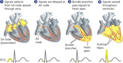
The heart's rhythmic contractions are coordinated by specialized cardiac muscle cells that generate and conduct electrical impulses. This system ensures that the atria and ventricles contract in the correct sequence.
Sinoatrial (SA) Node: Located in the right atrium, acts as the heart's pacemaker by initiating electrical impulses that set the heart rate.
Atrioventricular (AV) Node: Receives impulses from the SA node and delays them briefly, allowing the atria to contract and empty before the ventricles contract.
Bundle Branches and Purkinje Fibers: Conduct impulses rapidly throughout the ventricles, ensuring coordinated contraction.
Electrocardiogram (ECG): A recording of the electrical activity of the heart, used to assess heart rhythm and function.
Regulation of Heart Rate
The heart rate is regulated by physiological cues, including the nervous system, hormones, and body temperature:
Sympathetic Nervous System: Increases heart rate during activity or stress ("fight-or-flight" response).
Parasympathetic Nervous System: Decreases heart rate during rest.
Hormones: Epinephrine (adrenaline) increases heart rate.
Temperature: An increase in body temperature raises heart rate.
Summary Table: Key Structures and Functions in Mammalian Circulation
Structure | Function |
|---|---|
Right Atrium | Receives deoxygenated blood from the body |
Right Ventricle | Pumps deoxygenated blood to the lungs |
Left Atrium | Receives oxygenated blood from the lungs |
Left Ventricle | Pumps oxygenated blood to the body |
AV Valves | Prevent backflow into atria |
Semilunar Valves | Prevent backflow into ventricles |
SA Node | Pacemaker; initiates heart rhythm |
AV Node | Delays impulse, coordinates contraction |
Example: If a molecule of carbon dioxide starts in an arteriole in the right thumb and leaves the body in exhaled air, it passes through two capillary beds: one in the systemic circuit (body tissues) and one in the pulmonary circuit (lungs).
