Back
BackDNA and Its Role in Heredity: Structure, Replication, and Repair
Study Guide - Smart Notes
 Tailored notes based on your materials, expanded with key definitions, examples, and context.
DNA and Its Role in Heredity
Introduction
DNA (deoxyribonucleic acid) is the hereditary material in almost all living organisms. Its structure, replication, and repair mechanisms are central to understanding genetics and molecular biology. This chapter explores the experimental evidence for DNA as genetic material, its molecular structure, the process of replication, and the mechanisms that maintain its integrity.
Experiments Revealing DNA as Genetic Material
Early Evidence and Key Experiments
Chromosomes were known to contain both DNA and proteins, but it was unclear which was the genetic material.
DNA was found in the right place (chromosomes), in the right amounts, and varied among species, suggesting its role in heredity.
Griffith's Transformation Experiment
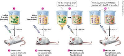
Two strains of Streptococcus pneumoniae were used: virulent (S) and nonvirulent (R).
Mixing heat-killed S strain with live R strain transformed the R strain into a virulent form, indicating a "transforming principle."
Avery, MacLeod, and McCarty's Experiment
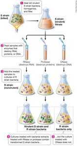
Identified DNA as the transforming principle by selectively destroying DNA, RNA, or protein in extracts from S cells.
Only destruction of DNA prevented transformation, confirming DNA as the genetic material.
Hershey-Chase Experiment
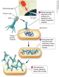
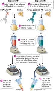
Used bacteriophage T2 to infect E. coli.
Phages were labeled with radioactive isotopes: 35S for protein, 32P for DNA.
After infection, only 32P (DNA) entered the bacteria, proving DNA carries genetic information.
Transfection and Transgenic Organisms
DNA can be introduced into eukaryotic cells (transfection), creating transgenic organisms.
Genetic markers (e.g., antibiotic resistance) are used to confirm successful transfection.
DNA Structure and Its Functional Implications
Chemical Composition and Base Pairing
DNA is a polymer of nucleotides, each containing a sugar, phosphate, and nitrogenous base.
Bases are classified as purines (adenine, guanine) and pyrimidines (cytosine, thymine).
Chargaff’s rule: Amount of purines equals pyrimidines (A = T, G = C), but A+T and G+C ratios vary among species.
Evidence from X-ray Diffraction
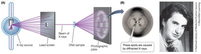
Rosalind Franklin’s X-ray crystallography revealed a helical structure with 10 nucleotides per turn and a diameter of 2 nm.
The Double Helix Model
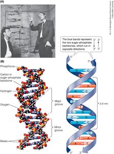
Watson and Crick built a model of DNA as a right-handed double helix with antiparallel strands.
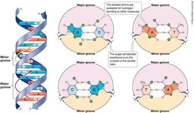
Complementary base pairing (A-T, G-C) ensures uniform width and accurate replication.
Sugar-phosphate backbones are on the outside; bases are on the inside.
Major and Minor Grooves
The double helix has major and minor grooves, exposing base pair edges for protein binding and regulation.
Key Features of DNA Structure
Double-stranded, right-handed helix
Antiparallel strands (5′ to 3′ and 3′ to 5′)
Hydrogen bonds between complementary bases
van der Waals forces between stacked bases
Accessible grooves for protein-DNA interactions
DNA Replication: The Semiconservative Model
Models of Replication
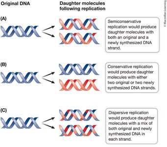
Semiconservative: Each new DNA molecule has one old and one new strand.
Conservative: Original molecule is conserved; new molecule is entirely new.
Dispersive: DNA fragments are mixed in each strand.
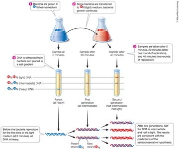
Meselson-Stahl Experiment
Used isotopes 15N and 14N to distinguish old and new DNA strands in E. coli.
Results supported the semiconservative model.
Steps in DNA Replication
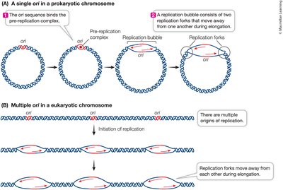
Initiation: Double helix unwinds at the origin of replication (ori).
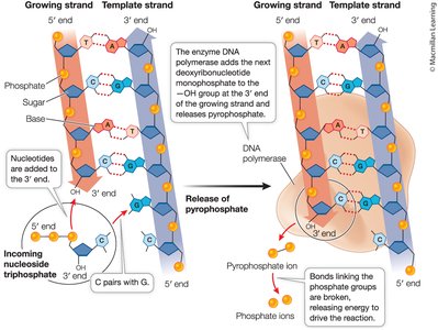
Elongation: DNA polymerase adds nucleotides to the 3′ end, forming phosphodiester bonds.
Termination: Replication ends when the entire molecule is copied.
Mechanism of DNA Synthesis
Nucleotides are added as deoxyribonucleoside triphosphates (dNTPs); two phosphates are released, providing energy for the reaction.
Origins of Replication
Prokaryotes have a single origin; eukaryotes have multiple origins to speed up replication.
Primers and DNA Polymerase
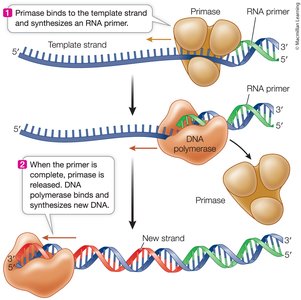
DNA polymerase requires a short RNA primer, synthesized by primase, to begin synthesis.
After elongation, the primer is removed and replaced with DNA.
Enzyme Structure and Function
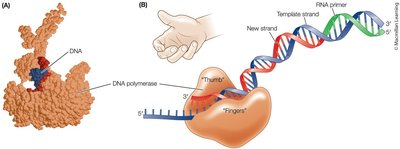
DNA polymerase has a "hand" shape: the palm is the active site, and the fingers recognize bases.
Other Proteins in Replication
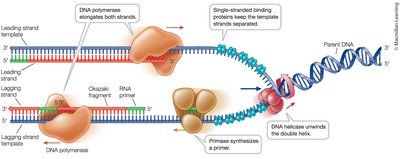
Helicase unwinds DNA; single-strand binding proteins stabilize unwound strands.
Leading and Lagging Strands
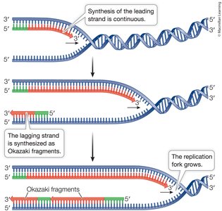
Leading strand is synthesized continuously toward the replication fork.
Lagging strand is synthesized discontinuously in Okazaki fragments, away from the fork.
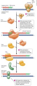
Joining Okazaki Fragments
DNA polymerase I replaces RNA primers with DNA; DNA ligase seals the fragments together.
Processivity and Sliding Clamp
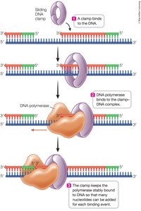
DNA polymerases are processive, adding many nucleotides per binding event.
A sliding DNA clamp protein keeps the polymerase attached to DNA.
Telomeres and Telomerase
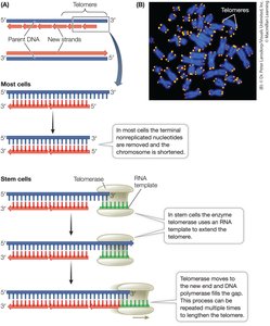
Telomeres are repetitive sequences at chromosome ends, protecting genes from loss during replication.
Telomerase extends telomeres in stem cells and cancer cells, allowing continued division.
DNA Repair Mechanisms
Types of DNA Repair
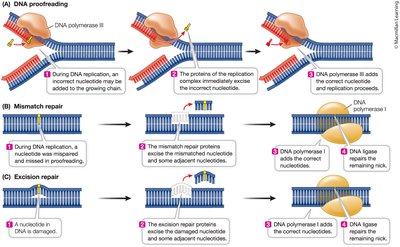
Proofreading: DNA polymerase removes mismatched bases during replication.
Mismatch repair: Proteins scan newly replicated DNA and correct errors missed by proofreading.
Excision repair: Enzymes remove damaged bases; DNA polymerase fills in the gap.
Targeting DNA Replication in Cancer Therapy
Cisplatin and DNA Cross-Linking
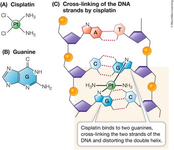
Cisplatin is a chemotherapy drug that forms covalent bonds with guanine bases, cross-linking DNA strands.
This cross-linking prevents DNA replication, leading to cell death (apoptosis), and is effective against certain cancers.