 Back
BackDNA and the Gene: Synthesis and Repair – Study Notes
Study Guide - Smart Notes
 Tailored notes based on your materials, expanded with key definitions, examples, and context.
DNA and the Gene: Synthesis and Repair
Testing Early Hypotheses about DNA Synthesis
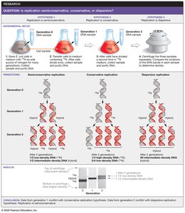
Understanding how DNA replicates was a foundational question in molecular biology. Three main hypotheses were proposed for DNA replication: semiconservative, conservative, and dispersive replication.
Semiconservative replication: Each daughter DNA molecule consists of one old (parental) strand and one newly synthesized strand.
Conservative replication: The parental DNA molecule remains intact, and an entirely new molecule is synthesized.
Dispersive replication: Parental DNA is cut into pieces, and each daughter molecule contains interspersed segments of old and new DNA.
Meselson–Stahl Experiment: This classic experiment used isotopic labeling to demonstrate that DNA replication is semiconservative.
A Model for DNA Synthesis
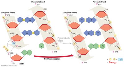
DNA synthesis is catalyzed by the enzyme DNA polymerase, which adds nucleotides to the 3' end of a growing DNA strand. DNA polymerases can only synthesize DNA in the 5' to 3' direction.
DNA polymerase: Enzyme responsible for DNA synthesis; several types exist in cells.
Directionality: DNA polymerase adds deoxyribonucleotides only to the 3' end, so synthesis always proceeds 5' → 3'.
Monomers: The building blocks are deoxyribonucleoside triphosphates (dNTPs), which have high potential energy due to their three phosphate groups. Hydrolysis of these phosphates drives the formation of phosphodiester bonds, making the reaction exergonic.
Where Does Replication Start?
DNA replication begins at specific locations called origins of replication, where the DNA double helix is unwound to form a replication bubble.
Origin of replication: Specific sequence where DNA synthesis is initiated.
Bacteria: Typically have a single origin and one replication bubble per chromosome.
Eukaryotes: Have multiple origins per chromosome, forming many replication bubbles.
Replication forks: Each bubble has two replication forks, and synthesis is bidirectional.

How is the Helix Opened and Stabilized?
Several proteins are required to open and stabilize the DNA double helix during replication:
DNA helicase: Breaks hydrogen bonds between DNA strands, separating them.
Single-strand DNA-binding proteins (SSBPs): Bind to separated strands to prevent them from re-annealing.
Topoisomerase: Relieves tension caused by unwinding by cutting and rejoining DNA downstream of the replication fork.
How Is the Leading Strand Synthesized?
Because DNA strands are antiparallel, the synthesis process differs for each strand. The leading strand is synthesized continuously toward the replication fork.
Primer: DNA polymerase cannot start synthesis de novo; it requires a short RNA primer with a free 3' end, synthesized by primase (an RNA polymerase).
Continuous synthesis: DNA polymerase adds dNTPs to the primer’s 3' end, synthesizing the leading strand in the 5' → 3' direction.

How is the Lagging Strand Synthesized?
The lagging strand is synthesized discontinuously, away from the replication fork, in short fragments called Okazaki fragments.
Discontinuous replication hypothesis: Primase synthesizes new RNA primers as the fork opens, and DNA polymerase synthesizes short DNA fragments.
Okazaki fragments: Short DNA segments synthesized on the lagging strand, later joined into a continuous strand by DNA ligase.


Proteins Required for DNA Synthesis in Bacteria
Multiple proteins coordinate the process of DNA replication. The following table summarizes their structure and function:
Protein | Structure | Function |
|---|---|---|
Helicase | Hexameric ring | Catalyzes the separation of DNA strands to open the double helix |
Single-strand DNA-binding proteins (SSBPs) | Tetramer | Stabilize single-stranded DNA |
Topoisomerase | Monomer | Breaks and rejoins the DNA double helix to relieve twisting forces |
Primase | Monomer | Catalyzes the synthesis of the RNA primer |
DNA polymerase III | Multisubunit complex | Extends the leading strand and Okazaki fragments |
Sliding clamp | Ring-shaped dimer | Holds DNA polymerase in place during strand extension |
DNA polymerase I | Monomer | Removes RNA primer and replaces it with DNA |
DNA ligase | Monomer | Catalyzes the joining of Okazaki fragments into a continuous strand |

The Replisome: DNA-Synthesizing Machine
The replisome is a large macromolecular complex containing all the enzymes required for DNA synthesis at the replication fork. It is dynamic, and recent observations suggest that the process is more variable than previously thought.

Replicating the Ends of Linear Chromosomes
Replication of linear eukaryotic chromosomes presents a unique challenge at the ends, known as telomeres.
Telomeres: Repetitive, non-coding DNA sequences at chromosome ends that protect genes from erosion during replication.
End replication problem: After removal of the final RNA primer on the lagging strand, DNA polymerase cannot fill in the gap, leading to progressive shortening of chromosomes with each cell division.

Telomerase Solves the End Replication Problem
Telomerase is an enzyme that extends telomeres, using an internal RNA template to add DNA repeats to the 3' end of the lagging strand template. This allows normal DNA polymerases to complete replication of chromosome ends.
Mechanism: Telomerase binds to the overhang, extends it, and allows primase and DNA polymerase to synthesize the complementary strand.
Cell types: Telomerase is active in gametes and stem cells, but not in most somatic cells.

Effect of Telomere Length on Cell Division
Telomere length limits the number of times a cell can divide. In most somatic cells, telomeres shorten with each division, eventually triggering cell senescence. In contrast, cancer cells often reactivate telomerase, enabling unlimited division.
Correcting Mistakes in DNA Synthesis
DNA polymerase has high fidelity, but errors can occur during replication. Several mechanisms ensure accuracy:
Proofreading: DNA polymerase has a 3' → 5' exonuclease activity that removes mismatched nucleotides immediately after they are added.
Mismatch repair: Specialized enzymes recognize and repair mismatches missed by DNA polymerase after replication is complete.

Repairing Damaged DNA
DNA can be damaged by environmental factors such as UV light, X-rays, and chemicals. Organisms have evolved repair systems to maintain genome integrity.
Thymine dimers: UV light can cause adjacent thymine bases to form covalent bonds, creating a kink that blocks replication.
Nucleotide excision repair: A protein complex recognizes distortions in the DNA helix, removes the damaged section, and fills in the gap using the undamaged strand as a template. DNA ligase seals the repaired strand.
Summary Table: Key Proteins in DNA Replication
Protein | Function |
|---|---|
Helicase | Unwinds DNA double helix |
SSBPs | Stabilize single-stranded DNA |
Topoisomerase | Relieves supercoiling tension |
Primase | Synthesizes RNA primers |
DNA polymerase III | Synthesizes new DNA strands |
DNA polymerase I | Removes RNA primers, fills gaps |
DNA ligase | Joins Okazaki fragments |
