 Back
DNA as the Genetic Material: Structure, Replication, and Packaging
Study Guide - Smart Notes
 Tailored notes based on your materials, expanded with key definitions, examples, and context.
DNA as the Genetic Material
Discovery of DNA as Genetic Material
Early 20th-century scientists debated whether proteins or DNA carried genetic information. Landmark experiments established DNA as the hereditary molecule, revolutionizing genetics and molecular biology.
Griffith's Transformation Experiment (1928): Demonstrated that a "transforming principle" from dead pathogenic bacteria could permanently change harmless bacteria into a pathogenic form, suggesting the transfer of genetic material.
Avery-MacLeod-McCarty Experiment (1944): Identified DNA as the transforming principle by showing that only destruction of DNA prevented transformation.
Hershey-Chase Experiment (1952): Used radioactive labeling to show that viral DNA, not protein, enters bacteria and directs viral replication, confirming DNA as the genetic material.
Conclusion: The ability of cells to take up and incorporate foreign DNA underlies genetic engineering and transformation techniques.
DNA Structure
Double Helix and Base Pairing
The elucidation of DNA's structure explained its capacity for information storage and faithful replication. DNA is a double helix composed of two antiparallel strands of nucleotides.
Nucleotide Structure: Each nucleotide contains a nitrogenous base (adenine, guanine, cytosine, or thymine), a deoxyribose sugar, and a phosphate group.
Chargaff's Rules: In any DNA, the amount of adenine (A) equals thymine (T), and guanine (G) equals cytosine (C), indicating complementary base pairing.
Rosalind Franklin's X-ray Crystallography: Revealed the helical structure and uniform width of DNA, critical for Watson and Crick's model.
Watson and Crick's Double Helix Model (1953): DNA consists of two antiparallel strands with a sugar-phosphate backbone and paired bases (A with T, G with C) forming the rungs of the ladder.
Base-Pairing Rules: A pairs with T via two hydrogen bonds; G pairs with C via three hydrogen bonds. This specificity ensures accurate replication and stability.
Mnemonic: "The Apple is Under the Tree" (A pairs with T); "The Car goes in the Garage" (C pairs with G).
Example: If a DNA sample contains 30% adenine, it must also contain 30% thymine. The remaining 40% is split equally between guanine and cytosine (20% each).
Complementary Strand Example: For a DNA sequence 5'-ATGC-3', the complementary strand is 3'-TACG-5'.
DNA Replication
Semi-Conservative Replication and Enzymatic Mechanisms
DNA replication is the process by which a cell copies its DNA, ensuring genetic continuity. The process is semi-conservative: each new DNA molecule contains one parental and one newly synthesized strand.
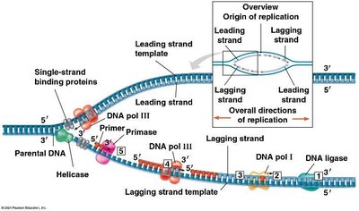
Origins of Replication: Replication begins at specific sites, forming replication bubbles with two replication forks.
Key Enzymes and Steps:
Helicase: Unwinds the DNA double helix.
Single-strand binding proteins: Stabilize unwound DNA strands.
Topoisomerase: Relieves strain ahead of the replication fork.
Primase: Synthesizes short RNA primers to initiate DNA synthesis.
DNA Polymerase III: Adds nucleotides to the 3' end of the primer or growing strand; proofreads new DNA.
DNA Polymerase I: Removes RNA primers and fills gaps with DNA.
DNA Ligase: Seals breaks in the sugar-phosphate backbone, joining Okazaki fragments.
Leading vs. Lagging Strands:
The leading strand is synthesized continuously toward the replication fork.
The lagging strand is synthesized discontinuously in short segments (Okazaki fragments) away from the fork; DNA ligase joins these fragments.
Example: The replication fork acts like a zipper: the leading strand is zipped smoothly, while the lagging strand is zipped in segments.
DNA Damage and Mutations
Sources and Repair of Mutations
Although DNA replication is highly accurate, errors and environmental factors can cause mutations—permanent changes in the DNA sequence. Mutations are essential for evolution but can also cause disease.
Causes of Mutations: Errors during replication, exposure to mutagens (chemicals), and high-energy radiation (UV, X-rays).
DNA Repair Mechanisms: Systems like nucleotide excision repair correct most errors.
Example: UV radiation can cause thymine dimers, distorting the DNA helix and potentially leading to skin cancer if unrepaired.
Telomeres
Protecting Chromosome Ends
Linear chromosomes in eukaryotes face end-replication problems. Telomeres—repetitive, non-coding sequences at chromosome ends—protect essential genes from erosion during replication.
Telomerase: An enzyme that extends telomeres in germ cells and some cancer cells, allowing continued division. In most somatic cells, telomerase is inactive, leading to gradual telomere shortening and cellular aging.
Example: Telomeres are like plastic tips on shoelaces, preventing fraying of the important genetic material.
DNA Packaging
Organization of DNA in Cells
To fit within the nucleus, DNA must be efficiently packaged. Packaging also regulates gene accessibility and expression.
Prokaryotes: DNA is a single circular chromosome, supercoiled in the nucleoid region.
Eukaryotes: Linear DNA wraps around histone proteins to form nucleosomes ("beads on a string"). Chromatin further coils to form compact chromosomes.
Euchromatin: Loosely packed, transcriptionally active.
Heterochromatin: Densely packed, transcriptionally inactive.
Example: DNA packaging is like winding a long string around spools (histones) and stacking them to fit in a small box (nucleus), with some sections easily accessible and others tightly locked away.
