 Back
DNA Fingerprinting and Genetics: Laboratory Study Guide
Study Guide - Smart Notes
 Tailored notes based on your materials, expanded with key definitions, examples, and context.
DNA Fingerprinting
Introduction to DNA Fingerprinting
DNA fingerprinting is a molecular technique used to identify individuals based on unique patterns in their DNA. This method is highly accurate and is widely used in forensic science, paternity testing, evolutionary biology, and the identification of individuals in various biological contexts.
Definition: DNA fingerprinting refers to the analysis of DNA fragment patterns to distinguish between individuals.
Applications: Forensic identification, paternity cases, identifying remains, evolutionary studies.
Sources of DNA: Blood, hair, semen, saliva, bone marrow, and other tissues.
Historical Methods: RFLP Analysis
Early DNA fingerprinting relied on Restriction Fragment Length Polymorphisms (RFLPs). This method uses restriction enzymes to cut DNA at specific sequences, generating fragments of varying lengths unique to each individual.
Restriction Enzymes: Proteins that recognize and cut DNA at specific nucleotide sequences (restriction sites).
Restriction Fragments: DNA pieces generated by enzyme digestion; their lengths vary among individuals due to sequence differences.
RFLPs: Variations in DNA fragment lengths caused by differences in restriction sites; serve as a genetic fingerprint.
Limitations: Requires large amounts of high-quality DNA and is time-consuming.
Modern Methods: PCR-Based STR/VNTR Analysis
Modern DNA fingerprinting uses the Polymerase Chain Reaction (PCR) to amplify regions of DNA with high variability, such as Short Tandem Repeats (STRs) and Variable Number Tandem Repeats (VNTRs).
PCR: A technique to make millions of copies of specific DNA regions from small or degraded samples.
STRs/VNTRs: DNA sequences where a short pattern is repeated; the number of repeats varies among individuals.
DNA Profile: The combined pattern of STRs/VNTRs at multiple locations, unique to each individual.
Gel Electrophoresis
Gel electrophoresis is used to separate DNA fragments by size. DNA samples are loaded into wells in an agarose gel and subjected to an electric field. DNA, being negatively charged, migrates toward the positive electrode. Smaller fragments move faster and farther than larger ones.
Agarose Gel: A porous matrix derived from algae, used to separate DNA fragments.
Electrophoresis Chamber: Apparatus where the gel is placed and an electric current is applied.
Direction of Movement: DNA moves toward the positive electrode due to its negative charge.
Band Visualization: After separation, DNA bands are visualized to determine fragment sizes and patterns.
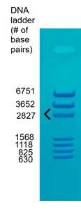
Example: The DNA ladder in the image above provides reference bands of known sizes (in base pairs) to estimate the sizes of sample DNA fragments.
Interpreting Band Patterns
The intensity of bands in a gel reflects the concentration of DNA fragments of a particular size. Multiple bands indicate the presence of fragments of different sizes, and their comparison is crucial for identification.
Band Intensity: Darker bands indicate higher concentrations of DNA fragments of that size.
Multiple Bands: Comparing several bands increases the accuracy of identification.
Laboratory Protocols
Protocols differ slightly depending on whether RFLP or PCR-based methods are used, but both involve preparing the gel, loading DNA samples, running the gel, and visualizing the results.
Sample Loading: Use a micropipette to load DNA samples into wells; avoid cross-contamination by changing tips between samples.
Running the Gel: Apply voltage and allow DNA to separate for a set time.
Visualization: Use a blue light box to view the separated DNA bands.

Example: The diagram above shows a gel with six wells, each for a different DNA sample, as used in the RFLP-based fingerprinting method.
Genetics Practice
Genetic Crosses and Punnett Squares
Punnett squares are used to predict the outcomes of genetic crosses. They show the possible combinations of alleles from the parents and the resulting genotypes and phenotypes of the offspring.
Allele: Different forms of a gene (e.g., B and b for body color in fruit flies).
Genotype: The genetic makeup of an organism (e.g., Bb, BB, or bb).
Phenotype: The observable trait (e.g., brown or black body color).
Dominance: Complete dominance, incomplete dominance, and codominance affect phenotype ratios.
Types of Inheritance
Complete Dominance: One allele completely masks the effect of the other (e.g., brown body color in fruit flies).
Incomplete Dominance: Heterozygotes show an intermediate phenotype (e.g., pink flowers from red and white alleles).
Codominance: Both alleles are fully expressed in heterozygotes (e.g., black and white feathers in chickens).
Sample Punnett Square Analysis
Fruit Fly Body Color (Bb x Bb): Brown is dominant (B), black is recessive (b).
Genotype Ratio: 1 BB : 2 Bb : 1 bb
Phenotype Ratio: 3 brown : 1 black
Example Calculation: For a cross between two heterozygotes (Bb x Bb):
Horses (AA x Aa): Smooth mane is dominant (A), curly is recessive (a). All offspring will have at least one A allele, so 100% will have a smooth mane.
Flowers (Rr x rr): Incomplete dominance; red (RR), pink (Rr), white (rr).
Genotype Ratio: 1 Rr : 1 rr
Phenotype Ratio: 1 pink : 1 white
Chickens (XX x XW): Codominance; black (XX), white (WW), black and white (XW).
Offspring with both colors: 50% (XW genotype)
Human Traits (Mid-digital Hair): Dominant allele (M) for hair, recessive (m) for no hair. If one parent is heterozygous (Mm) and the other is homozygous recessive (mm), 50% chance for mid-digital hair in offspring.
Cystic Fibrosis: Recessive trait (f). If one parent is ff (has disease) and the other is Ff (carrier), 50% chance for child to have cystic fibrosis.
Combined Traits (Cystic Fibrosis and Huntington's Disease): Use the FOIL method and a dihybrid Punnett square to determine the probability of inheriting both traits.
Structure of Nucleic Acids
Nucleotide: The building block of DNA and RNA, consisting of three parts:
Phosphate group
Pentose sugar (deoxyribose in DNA, ribose in RNA)
Nitrogenous base (A, T, C, G in DNA; A, U, C, G in RNA)
Comparison of DNA and RNA
Feature | DNA | RNA |
|---|---|---|
Sugar | Deoxyribose | Ribose |
Bases | A, T, C, G | A, U, C, G |
Strands | Double-stranded | Single-stranded |
Function | Genetic information storage | Protein synthesis, gene regulation |
Additional info: In DNA fingerprinting, comparing multiple bands (loci) increases the statistical power of identification, making it extremely unlikely for two unrelated individuals to share the same DNA profile. In genetics, understanding inheritance patterns is crucial for predicting trait distribution and for medical genetics applications.
