Back
DNA Replication and Gene Expression: Structure, Mechanisms, and Regulation
Study Guide - Smart Notes
 Tailored notes based on your materials, expanded with key definitions, examples, and context.
DNA Structure and Organization
DNA as the Genetic Material
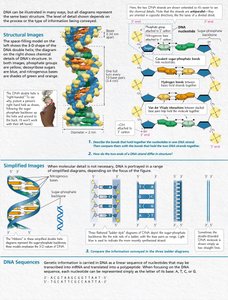
DNA (deoxyribonucleic acid) is the hereditary material in almost all living organisms, encoding the instructions for cellular structure and function. Its double helix structure allows for the precise storage and transmission of genetic information.
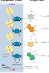
Nucleotide: The basic unit of DNA, consisting of a phosphate group, deoxyribose sugar, and a nitrogenous base (adenine, thymine, cytosine, or guanine).
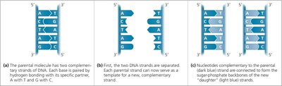
Double Helix: Two antiparallel strands held together by hydrogen bonds between complementary bases (A-T, G-C).
Chromatin: DNA is packaged with proteins (histones) into chromatin, which can be further organized into chromosomes.
Chromatin Structure: Euchromatin and Heterochromatin
Chromatin exists in two forms: euchromatin (less condensed, transcriptionally active) and heterochromatin (highly condensed, transcriptionally inactive). DNA wraps around histone proteins to form nucleosomes, the basic unit of chromatin structure.
Euchromatin: Loosely packed, accessible for transcription.
Heterochromatin: Densely packed, generally not transcribed.
DNA Replication
Overview and Models of Replication
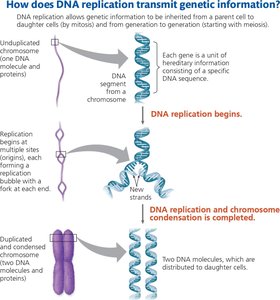
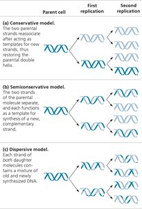
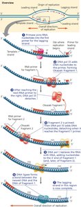
DNA replication is the process by which a cell copies its DNA before cell division. The semiconservative model describes how each new DNA molecule consists of one parental and one newly synthesized strand.
Semiconservative Replication: Each daughter DNA molecule contains one old and one new strand.
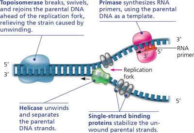
Replication Fork: The Y-shaped region where DNA is unwound and new strands are synthesized.
Enzymes and Proteins in DNA Replication
Multiple enzymes coordinate the accurate and efficient replication of DNA:
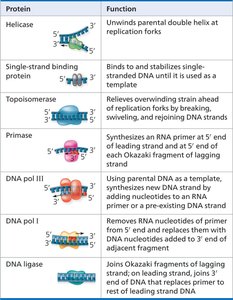
Helicase: Unwinds the DNA double helix at the replication fork.
Single-Strand Binding Proteins: Stabilize unwound DNA strands.
Topoisomerase: Relieves strain ahead of the replication fork by breaking and rejoining DNA strands.
Primase: Synthesizes short RNA primers needed for DNA polymerase to begin synthesis.
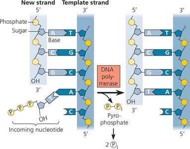
DNA Polymerase III: Adds nucleotides to the 3' end of the primer, synthesizing new DNA.
DNA Polymerase I: Removes RNA primers and replaces them with DNA.
DNA Ligase: Joins Okazaki fragments on the lagging strand.
Leading and Lagging Strands
DNA polymerase can only synthesize DNA in the 5' to 3' direction. This results in continuous synthesis on the leading strand and discontinuous synthesis (Okazaki fragments) on the lagging strand.
Leading Strand: Synthesized continuously toward the replication fork.
Lagging Strand: Synthesized discontinuously away from the fork in short segments called Okazaki fragments.
DNA Repair and Telomeres
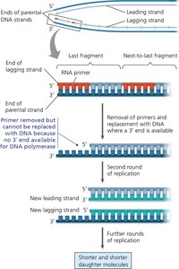
Cells possess mechanisms to repair DNA damage and maintain genome integrity. Telomeres protect chromosome ends but shorten with each replication cycle in most somatic cells.
Mismatch Repair: Corrects errors missed by DNA polymerase proofreading.
Nucleotide Excision Repair: Removes and replaces damaged DNA segments.
Telomeres: Repetitive DNA sequences at chromosome ends that prevent loss of genetic information.
Gene Expression: From DNA to Protein
Central Dogma: Transcription and Translation
The central dogma of molecular biology describes the flow of genetic information from DNA to RNA (transcription) and from RNA to protein (translation).
Transcription: Synthesis of RNA from a DNA template.
Translation: Synthesis of a polypeptide using the information in mRNA.
Codon: A three-nucleotide sequence in mRNA that specifies an amino acid.
Transcription: Initiation, Elongation, and Termination
Transcription involves three main steps: initiation (RNA polymerase binds to the promoter), elongation (RNA strand is synthesized), and termination (RNA polymerase releases the completed RNA transcript).
Promoter: DNA sequence where RNA polymerase binds to initiate transcription.
RNA Polymerase: Enzyme that synthesizes RNA from the DNA template.
Terminator: Sequence signaling the end of transcription (in prokaryotes).
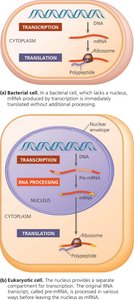
RNA Processing in Eukaryotes
In eukaryotes, primary RNA transcripts (pre-mRNA) undergo processing before becoming mature mRNA:
5' Cap: Modified guanine nucleotide added to the 5' end.
Poly-A Tail: String of adenine nucleotides added to the 3' end.
RNA Splicing: Removal of noncoding introns and joining of exons.
Spliceosome: Complex responsible for RNA splicing, sometimes aided by ribozymes (RNA molecules with catalytic activity).
Translation: The Genetic Code and Protein Synthesis
Translation occurs at the ribosome, where tRNA molecules bring amino acids corresponding to mRNA codons. The process involves initiation, elongation, and termination phases.
tRNA: Adapter molecule with an anticodon that pairs with mRNA codons and carries specific amino acids.
Ribosome: Molecular machine composed of rRNA and proteins, facilitating peptide bond formation.
Wobble: Flexibility in base pairing at the third codon position, allowing some tRNAs to recognize multiple codons.
Gene Mutations and Phenotypic Effects
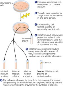
Mutations in DNA can alter gene expression and protein function, leading to observable changes in phenotype. For example, a single base change can result in a nonfunctional enzyme and a dramatic change in appearance.
Summary Table: Key Proteins in DNA Replication
Protein | Function |
|---|---|
Helicase | Unwinds parental double helix at replication forks |
Single-strand binding protein | Binds to and stabilizes single-stranded DNA until it is used as a template |
Topoisomerase | Relieves overwinding strain ahead of replication forks by breaking, swiveling, and rejoining DNA strands |
Primase | Synthesizes an RNA primer at 5' end of leading strand and at 5' end of each Okazaki fragment of lagging strand |
DNA pol III | Using parental DNA as template, synthesizes new DNA strand by covalently adding nucleotides to the 3' end of a pre-existing DNA strand or RNA primer |
DNA pol I | Removes RNA nucleotides of primer from 5' end and replaces them with DNA nucleotides added to 3' end of adjacent fragment |
DNA ligase | Joins Okazaki fragments of lagging strand; on leading strand, joins 3' end of DNA that replaces primer to rest of leading strand DNA |