 Back
DNA Replication: Structure, Mechanism, and Enzymatic Processes
Study Guide - Smart Notes
 Tailored notes based on your materials, expanded with key definitions, examples, and context.
DNA Structure and Discovery
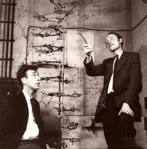
Watson and Crick's Double Helix Model
The structure of DNA was elucidated by James Watson and Francis Crick in 1953, who proposed the double helix model. Their discovery revealed how genetic information is stored and replicated in living organisms.
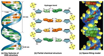
Double Helix: DNA consists of two strands coiled around each other, forming a helical structure.
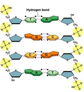
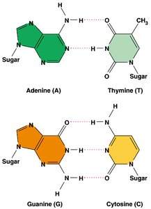
Base Pairing: The strands are held together by specific pairing between nitrogenous bases: adenine (A) pairs with thymine (T), and guanine (G) pairs with cytosine (C).
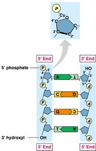
Antiparallel Orientation: The two strands run in opposite directions, one from 5' to 3' and the other from 3' to 5'.
Significance: The specific base pairing suggests a mechanism for copying genetic material during cell division.
DNA Structure: Chemical Composition
Nucleotides and the DNA Backbone
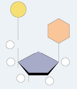
DNA is a polymer made up of repeating units called nucleotides. Each nucleotide consists of a phosphate group, a deoxyribose sugar, and a nitrogenous base.
Nitrogenous Bases: Purines (adenine and guanine) and pyrimidines (thymine and cytosine).
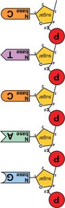
Sugar-Phosphate Backbone: Nucleotides are joined by phosphodiester bonds between the 3' hydroxyl group of one sugar and the 5' phosphate of the next.
Directionality: The backbone has directionality, with a 5' end (phosphate group) and a 3' end (hydroxyl group).
Base Pairing and Bonding
The two DNA strands are held together by hydrogen bonds between complementary bases. Covalent phosphodiester bonds form the backbone, while hydrogen bonds provide specificity and allow strand separation during replication.
A:T Pair: Two hydrogen bonds.
G:C Pair: Three hydrogen bonds (stronger pairing).
Mechanism of DNA Replication
Semi-Conservative Replication
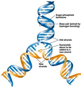
DNA replication is semi-conservative: each new DNA molecule consists of one parental strand and one newly synthesized strand. Base pairing allows each strand to serve as a template for the synthesis of a complementary strand.
Template Mechanism: Parental strands separate and serve as templates for new strands.
Result: Two DNA molecules, each with one old and one new strand.
Enzymes Involved in DNA Replication
DNA replication is a highly coordinated process involving several enzymes:
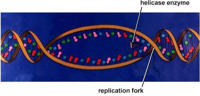
Helicase: Unwinds the DNA double helix at the replication fork.
Single-Stranded Binding Proteins (SSB): Stabilize unwound DNA strands.
Primase: Synthesizes short RNA primers to provide a starting point for DNA synthesis.
DNA Polymerase III: Main enzyme that adds nucleotides to the growing DNA strand in the 5' to 3' direction.
DNA Polymerase I: Removes RNA primers and replaces them with DNA nucleotides.
Ligase: Joins Okazaki fragments on the lagging strand, sealing nicks in the sugar-phosphate backbone.
Energy for DNA Synthesis
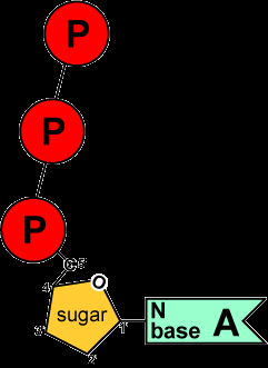
Nucleotides used in DNA synthesis are nucleoside triphosphates (e.g., dATP, dGTP, dCTP, dTTP). The energy for forming phosphodiester bonds comes from the hydrolysis of the high-energy phosphate bonds in these molecules.
Energy Source: The removal of two phosphate groups (pyrophosphate) provides the energy needed for polymerization.
Enzyme: DNA polymerase catalyzes the addition of nucleotides using this energy.
Directionality and Leading/Lagging Strands
DNA polymerase can only add nucleotides to the 3' end of a growing DNA strand. This leads to continuous synthesis on the leading strand and discontinuous synthesis on the lagging strand (Okazaki fragments).
Leading Strand: Synthesized continuously in the direction of the replication fork.
Lagging Strand: Synthesized discontinuously, forming short Okazaki fragments that are later joined by ligase.
Primer Requirement: DNA polymerase requires a primer with a free 3' OH group to initiate synthesis.
Replication Fork and Bubble
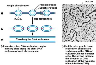
Replication begins at origins of replication, forming replication bubbles. Multiple replication forks allow rapid and efficient copying of large eukaryotic chromosomes.
Replication Fork: The Y-shaped region where DNA is actively being unwound and replicated.
Replication Bubble: The region of DNA where replication is occurring, with two replication forks moving in opposite directions.
Proofreading and Accuracy
Editing and Proofreading by DNA Polymerases
DNA polymerases possess proofreading activity, which ensures high fidelity during DNA replication. Errors are corrected by removing mismatched nucleotides and replacing them with the correct ones.
DNA Polymerase I: Removes RNA primers, repairs mismatches, and edits errors.
Error Rate: Proofreading reduces the error rate from 1 in 10,000 to 1 in 100 million bases.
Telomeres and Chromosome Erosion
End Replication Problem and Telomerase
During replication, the ends of linear chromosomes (telomeres) cannot be fully replicated, leading to gradual shortening with each cell division. Telomerase is an enzyme that extends telomeres, maintaining chromosome integrity in certain cells (e.g., stem cells, cancer cells).
Telomeres: Repetitive, non-coding sequences at chromosome ends that protect genetic information.
Telomerase: Adds telomeric repeats to the 5' ends, counteracting shortening.
Cellular Aging: Telomere shortening limits the number of cell divisions (Hayflick limit).
Summary Table: Key Enzymes in DNA Replication
Enzyme | Function |
|---|---|
Helicase | Unwinds DNA double helix |
Single-Stranded Binding Proteins | Stabilize unwound DNA |
Primase | Synthesizes RNA primers |
DNA Polymerase III | Main DNA synthesis enzyme |
DNA Polymerase I | Removes RNA primers, edits, and repairs DNA |
Ligase | Joins Okazaki fragments |
Telomerase | Extends telomeres |
Replication Speed and Accuracy
DNA replication is both rapid and highly accurate. For example, Escherichia coli can replicate its 5 million base pair genome in less than an hour, while human cells replicate 6 billion base pairs in a few hours with remarkable fidelity.
Speed: DNA polymerase III adds up to 1000 bases per second in prokaryotes.
Accuracy: Only about 1 error per 100 million bases, or ~30 errors per cell cycle in humans.
