 Back
DNA Structure and Replication: Discovery, Mechanisms, and Key Experiments
Study Guide - Smart Notes
 Tailored notes based on your materials, expanded with key definitions, examples, and context.
DNA Structure and Replication
Discovery of DNA as Genetic Material
The identification of DNA as the hereditary material was a pivotal moment in biology, established through a series of classic experiments that distinguished DNA from proteins as the carrier of genetic information.
Spontaneous Generation vs. Germ Theory: Early debates about the origin of life and disease transmission were resolved by experiments such as those by Louis Pasteur, who disproved spontaneous generation and supported the germ theory of disease.
Griffith-Avery Experiment (1928): Demonstrated that a 'transforming principle' from dead pathogenic bacteria could genetically alter harmless bacteria, suggesting the presence of a transferable genetic material.
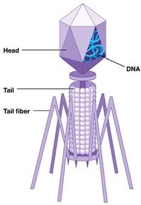
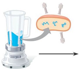
Hershey-Chase Experiment (1952): Used bacteriophages (viruses that infect bacteria) to show that DNA, not protein, is the genetic material. Radioactive isotopes labeled DNA and protein separately, revealing that only DNA entered bacterial cells and directed viral replication.
Structure of DNA
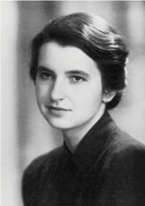
The structure of DNA was elucidated through the combined efforts of several scientists, culminating in the double helix model proposed by Watson and Crick, with critical contributions from Rosalind Franklin's X-ray diffraction data.
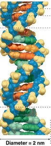
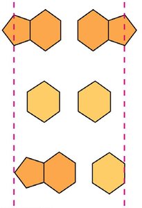
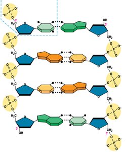
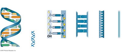
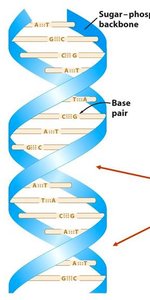
Double Helix: DNA consists of two antiparallel strands forming a right-handed double helix, with a sugar-phosphate backbone on the outside and nitrogenous bases paired in the interior.
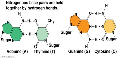
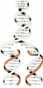
Base Pairing: Adenine (A) pairs with Thymine (T), and Guanine (G) pairs with Cytosine (C), held together by hydrogen bonds. This complementary base pairing is essential for accurate replication.
Major and Minor Grooves: The helical structure creates alternating major and minor grooves, which are important for protein-DNA interactions.
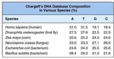
Chargaff’s Rules: The amount of A equals T, and G equals C in DNA, supporting the base-pairing model.
Key Features of DNA Structure
Double-stranded helix
Antiparallel orientation (5' to 3' and 3' to 5')
Right-handed helix
Major and minor grooves
DNA Packaging in Chromosomes
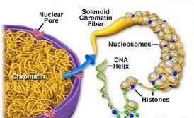
To fit within the microscopic nucleus, eukaryotic DNA is highly compacted through association with proteins called histones, forming chromatin and further condensing into chromosomes during cell division.
Nucleosome: The basic unit of DNA packaging, consisting of DNA wrapped around a histone core.
Chromatin: The complex of DNA and proteins that forms chromosomes.
Levels of Compaction: DNA wraps around nucleosomes ('beads on a string'), which coil into solenoid fibers and further condense during mitosis.

DNA Replication: Mechanisms and Models
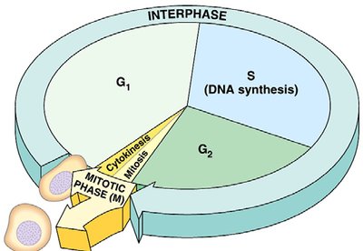
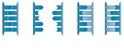
DNA replication is a highly regulated process ensuring the accurate duplication of genetic material before cell division. It follows the semiconservative model, where each new DNA molecule consists of one parental and one newly synthesized strand.
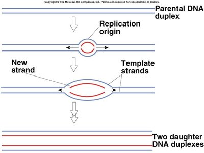
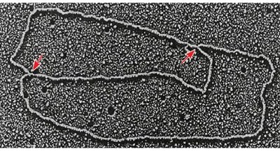
Origin of Replication (ORI): Specific sequence where replication begins. Prokaryotes have a single ORI; eukaryotes have multiple ORIs per chromosome.
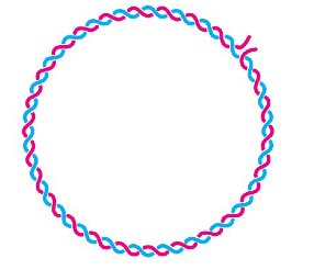
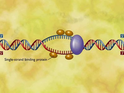
Replication Bubble and Fork: DNA unwinds at the ORI, forming a bubble with two replication forks where synthesis occurs bidirectionally.
Enzymes Involved:
Helicase: Unwinds the DNA double helix.
Single-Strand Binding Proteins (SSBP): Stabilize unwound DNA.
Topoisomerase: Relieves supercoiling ahead of the replication fork.
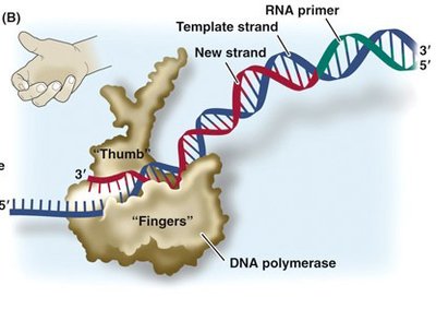
Primase: Synthesizes short RNA primers to initiate DNA synthesis.
DNA Polymerase: Adds nucleotides to the growing DNA strand in the 5' to 3' direction; requires a primer and a template.
Ligase: Seals nicks between Okazaki fragments on the lagging strand.
Leading vs. Lagging Strand:
Leading strand: Synthesized continuously toward the replication fork.
Lagging strand: Synthesized discontinuously away from the fork in short Okazaki fragments.
Limitations and Solutions in DNA Replication
End-Replication Problem: DNA polymerase cannot fully replicate the 5' ends of linear chromosomes, leading to progressive shortening.
Telomeres: Repetitive DNA sequences at chromosome ends that protect coding regions from loss. Telomerase extends telomeres in certain cells.
Proofreading and Repair: DNA polymerases have proofreading activity, and additional repair mechanisms (e.g., mismatch repair, nucleotide excision repair) correct errors, resulting in a very low mutation rate.
Key Terms and Enzymes
Spontaneous generation: Disproven hypothesis that life arises from non-living matter.
Replication: The process of copying DNA before cell division.
Germ theory: The theory that microorganisms cause disease.
Polymerize: To form a polymer by linking monomers.
Pathogenic: Capable of causing disease.
Nucleosome: DNA wrapped around histone proteins.
Chromatin: DNA-protein complex in eukaryotic cells.
Chromosome: Condensed chromatin visible during cell division.
Origin of replication (ORI): Site where DNA replication begins.
Bacteriophage: Virus that infects bacteria.
Okazaki fragment: Short DNA fragment synthesized on the lagging strand.
DNA polymerase: Enzyme that synthesizes DNA.
Single-strand binding protein: Stabilizes unwound DNA.
Helicase: Unwinds DNA helix.
Telomerase: Extends telomeres.
Primase: Synthesizes RNA primer.
Topoisomerase: Relieves supercoiling.
Ligase: Seals DNA nicks.
Table: Chargaff's DNA Composition in Various Species
Species | A (%) | T (%) | G (%) | C (%) |
|---|---|---|---|---|
Homo sapiens (human) | 31.0 | 31.5 | 19.1 | 18.4 |
Drosophila melanogaster (fruit fly) | 27.3 | 27.6 | 22.5 | 22.5 |
Zea mays (corn) | 25.6 | 25.3 | 24.5 | 24.6 |
Neurospora crassa (fungus) | 23.0 | 23.3 | 27.1 | 26.6 |
Escherichia coli (bacterium) | 24.6 | 24.3 | 25.5 | 25.6 |
Bacillus subtilis (bacterium) | 28.4 | 29.0 | 21.0 | 21.6 |
Key Equations and Concepts
Base Pairing Rule: $[A] = [T],\ [G] = [C]$
Semiconservative Replication: Each daughter DNA molecule contains one parental and one new strand.
Direction of Synthesis: DNA polymerase synthesizes DNA in the 5' to 3' direction only.
Summary Table: Leading vs. Lagging Strand Synthesis
Feature | Leading Strand | Lagging Strand |
|---|---|---|
Synthesis | Continuous | Discontinuous (Okazaki fragments) |
Direction | Toward replication fork | Away from replication fork |
Primer Requirement | One primer | Multiple primers |
Role of DNA Ligase | Minimal | Joins Okazaki fragments |
Example: Hershey-Chase Experiment
Radioactive phosphorus (32P) was used to label DNA, and radioactive sulfur (35S) was used to label protein. After infection of bacteria by phages, only 32P was found inside the cells, confirming DNA as the genetic material.
Additional info:
DNA repair mechanisms, such as mismatch repair and nucleotide excision repair, are essential for maintaining genetic stability and preventing mutations that could lead to disease.
Telomerase activity is high in germ cells and cancer cells, allowing for continued cell division without loss of genetic information at chromosome ends.
