 Back
BackDNA Structure and Replication: Foundations of Genetic Inheritance
Study Guide - Smart Notes
 Tailored notes based on your materials, expanded with key definitions, examples, and context.
DNA Structure and Replication
Introduction to Genetic Material
The genetic material of living organisms is DNA (deoxyribonucleic acid), which encodes the instructions for inheritance and cellular function. Understanding DNA's structure and replication is fundamental to molecular biology and genetics.
Discovery and Evidence for DNA Structure
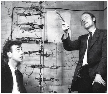
Historical Experiments: The double helix structure of DNA was elucidated through a combination of chemical analysis, X-ray diffraction, and model building.
X-ray Diffraction: X-ray images provided critical evidence for the helical structure of DNA.

Chemical Composition of DNA
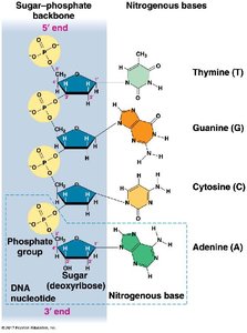
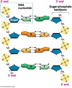
Nucleotides: DNA is a polymer of nucleotides, each consisting of a phosphate group, a deoxyribose sugar, and a nitrogenous base.
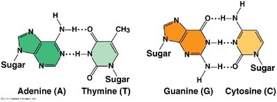
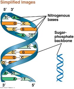
Nitrogenous Bases: The four bases are adenine (A), thymine (T), guanine (G), and cytosine (C).
Base Pairing: Adenine pairs with thymine (A-T) via two hydrogen bonds, and guanine pairs with cytosine (G-C) via three hydrogen bonds.
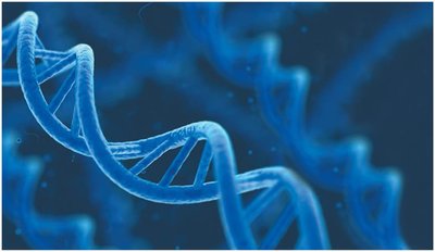
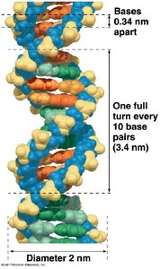
Double Helix Structure
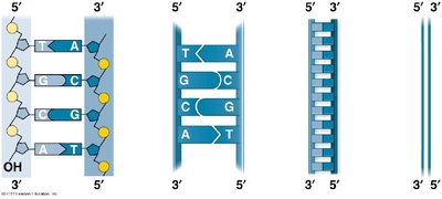
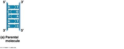
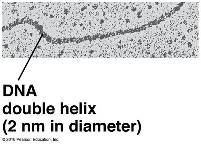
The DNA molecule consists of two antiparallel strands forming a right-handed double helix. The sugar-phosphate backbones are on the outside, and the nitrogenous bases pair in the interior.
Antiparallel Orientation: One strand runs 5' to 3', the other 3' to 5'.
Helical Parameters: The helix has a diameter of 2 nm, with bases stacked 0.34 nm apart, and one full turn every 10 base pairs (3.4 nm).
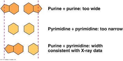
Base Pairing Rules and Consistency with X-ray Data
Purines and Pyrimidines: Purines (A, G) pair with pyrimidines (T, C) to maintain a uniform helix width.
Incorrect Pairings: Purine-purine pairs are too wide; pyrimidine-pyrimidine pairs are too narrow.
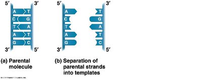
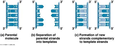
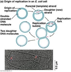
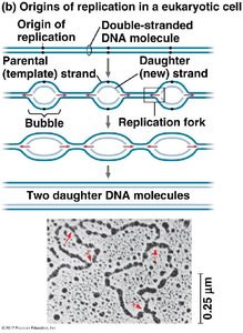
DNA Replication: The Semi-Conservative Model
DNA replication is the process by which a cell copies its DNA before cell division. The semi-conservative model states that each new DNA molecule consists of one parental and one newly synthesized strand.
Steps of Replication:
Parental DNA molecule unwinds.
Each strand serves as a template for a new complementary strand.
Result: Two DNA molecules, each with one old and one new strand.
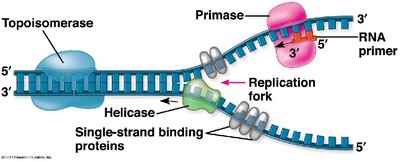
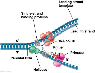
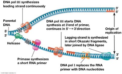
Mechanism of DNA Replication
Enzymes Involved:
Helicase: Unwinds the DNA double helix.
Single-Strand Binding Proteins: Stabilize unwound DNA.
Primase: Synthesizes RNA primers.
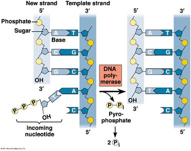
DNA Polymerase III: Extends new DNA strand from primer.
DNA Polymerase I: Replaces RNA primers with DNA.
DNA Ligase: Joins Okazaki fragments on the lagging strand.
Topoisomerase: Relieves supercoiling ahead of the replication fork.
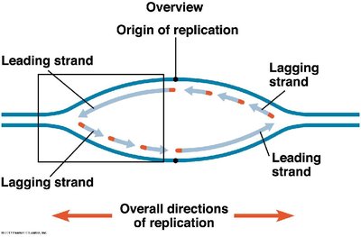
Leading and Lagging Strands:
Leading strand synthesized continuously in 5' to 3' direction.
Lagging strand synthesized discontinuously as Okazaki fragments.
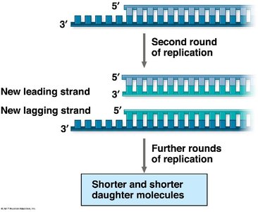
Replication at the Ends of Linear Chromosomes (Telomeres)
Linear eukaryotic chromosomes face the end-replication problem, where the lagging strand cannot be fully replicated, leading to progressive shortening of chromosomes with each cell division. Telomeres, repetitive DNA sequences at chromosome ends, and the enzyme telomerase help mitigate this loss.
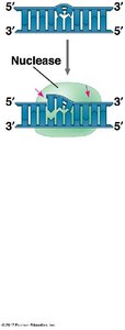
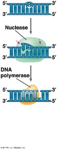
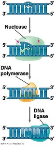
DNA Repair Mechanisms
Cells possess multiple mechanisms to repair damaged DNA, many of which utilize enzymes also involved in replication. The main steps include recognition and removal of damaged DNA, synthesis of replacement DNA, and sealing of the strand.
Nuclease: Removes damaged DNA segments.
DNA Polymerase: Fills in the gap with new nucleotides.
DNA Ligase: Seals the final nick in the sugar-phosphate backbone.
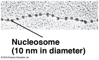
Packaging of DNA in Eukaryotic Cells
In eukaryotes, DNA is highly compacted to fit within the nucleus. DNA wraps around histone proteins to form nucleosomes, which further coil and fold to produce higher-order chromatin structures.
DNA Double Helix: 2 nm in diameter.
Nucleosome: DNA wrapped around histone core, 10 nm in diameter.
Structure | Diameter | Description |
|---|---|---|
DNA double helix | 2 nm | Basic structure of DNA |
Nucleosome | 10 nm | DNA wrapped around histone proteins |
Additional info: Higher-order chromatin fibers (30 nm, 300 nm, and metaphase chromosome) are formed by further folding and compaction, essential for chromosome segregation during cell division.
