 Back
BackDNA Synthesis: Experiments, Structure, and Replication
Study Guide - Smart Notes
 Tailored notes based on your materials, expanded with key definitions, examples, and context.
DNA Synthesis
Griffith's Experiment and Bacterial Transformation
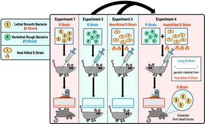
Frederick Griffith's 1928 experiment demonstrated that bacteria can transfer genetic material through a process called transformation. This finding was foundational in identifying DNA as the genetic material.
Transformation: The uptake of external DNA by a cell, resulting in a change in genotype and phenotype.
Griffith used two strains of Streptococcus pneumoniae: the smooth (S) strain (lethal, with a polysaccharide capsule) and the rough (R) strain (non-lethal, lacking the capsule).
When mice were injected with a mixture of heat-killed S strain and living R strain, the mice died, and living S strain bacteria were recovered, indicating that genetic material from the dead S strain had transformed the R strain into a pathogenic form.
The Hershey-Chase Experiment
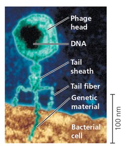
In 1952, Martha Hershey and Alfred Chase used bacteriophages (viruses that infect bacteria) to confirm that DNA, not protein, is the genetic material.
Bacteriophage: A virus that infects bacteria, consisting of a protein coat and a nucleic acid core (DNA or RNA).
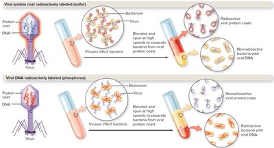
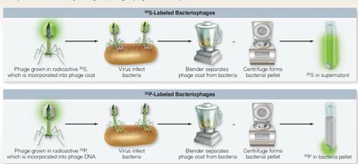
Hershey and Chase labeled viral DNA with radioactive phosphorus (32P) and viral protein with radioactive sulfur (35S).
After infection, only the radioactive DNA entered the bacterial cells, while the protein remained outside, confirming that DNA carries genetic information.
Chargaff's Rules
Erwin Chargaff discovered two key rules about DNA base composition:
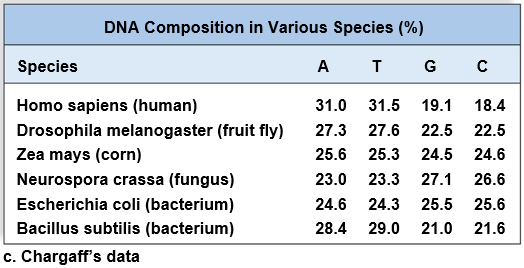
The proportion of the four nucleotide bases (A, T, G, C) varies between species.
Within a species, the amount of adenine (A) is approximately equal to thymine (T), and the amount of guanine (G) is approximately equal to cytosine (C).
Species | A | T | G | C |
|---|---|---|---|---|
Homo sapiens (human) | 31.0 | 31.5 | 19.1 | 18.4 |
Drosophila melanogaster (fruit fly) | 27.3 | 27.6 | 22.5 | 22.5 |
Zea mays (corn) | 25.6 | 25.3 | 24.5 | 24.6 |
Neurospora crassa (fungus) | 23.0 | 23.3 | 27.1 | 26.6 |
Escherichia coli (bacterium) | 24.6 | 24.3 | 25.5 | 25.6 |
Bacillus subtilis (bacterium) | 28.4 | 29.0 | 21.0 | 21.6 |
Discovery of DNA Structure
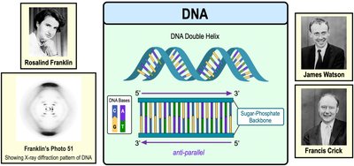
Rosalind Franklin used X-ray diffraction to capture images of DNA, which helped James Watson and Francis Crick deduce the double-helix structure of DNA in 1953.
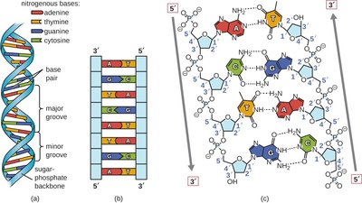
DNA is a double helix with two antiparallel strands of nucleotides.
Nucleotides on opposite strands pair via hydrogen bonds: A with T, and C with G (complementary base pairing).
Detailed DNA Structure
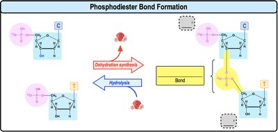
DNA consists of two strands of nucleotide monomers linked by phosphodiester bonds. Each strand has a 5' phosphate group at one end and a 3' hydroxyl group at the other.
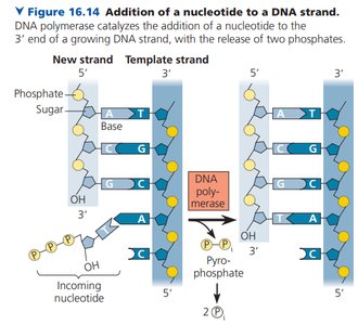
New nucleotides are always added to the 3' hydroxyl group of the existing DNA strand.
Complementary base pairing ensures a uniform width for the double helix and allows one strand to serve as a template for the other.
Meselson-Stahl Experiment: Semi-Conservative Replication
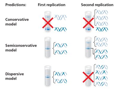
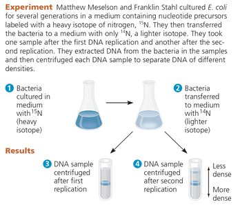
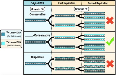
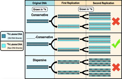
Matthew Meselson and Franklin Stahl demonstrated that DNA replication is semi-conservative: each new DNA molecule consists of one old (parental) strand and one newly synthesized strand.
Old strands serve as templates for new DNA synthesis.
Experiments using isotopes of nitrogen showed that after replication, DNA molecules contained one old and one new strand.
Introduction to DNA Replication
DNA replication is better understood in prokaryotes, but the process is fundamentally similar in eukaryotes. Parental strands separate and serve as templates for new DNA synthesis.
Replication requires several specialized enzymes and proteins, including topoisomerase, helicase, single-stranded binding proteins, primase, DNA polymerases, and DNA ligase.
Protein | Function |
|---|---|
Helicase | Unwinds parental double helix at replication forks |
Single-strand binding protein | Binds to and stabilizes single-stranded DNA until it is used as a template |
Topoisomerase | Relieves overwinding strain ahead of replication forks by breaking, swiveling, and rejoining DNA strands |
Primase | Synthesizes an RNA primer at 5' end of leading strand and at 5' end of each Okazaki fragment of lagging strand |
DNA pol III | Using parental DNA as a template, synthesizes new DNA strand by adding nucleotides to an RNA primer or a pre-existing DNA strand |
DNA pol I | Removes RNA nucleotides of primer from 5' end and replaces them with DNA nucleotides |
DNA ligase | Joins Okazaki fragments of lagging strand; on leading strand, joins 3' end of DNA that replaces primer to rest of leading strand DNA |
Origin of Replication and Replication Forks
DNA replication begins at specific sequences called origins of replication (ORI). Prokaryotes have circular chromosomes with a single ORI, while eukaryotes have linear chromosomes with multiple ORIs.
Proteins bind to the ORI and separate the two DNA strands, forming a replication bubble with Y-shaped replication forks at each end.
Replication proceeds bidirectionally from the ORI.
Unwinding the DNA: Topoisomerase, Helicase, and SSBs
Several proteins are involved in unwinding DNA during replication:
Topoisomerase: Relieves strain caused by supercoiling ahead of the replication fork.
Helicase: Unwinds DNA by breaking hydrogen bonds between bases, creating single-stranded DNA.
Single-Strand Binding Proteins (SSBs): Stabilize single-stranded DNA and prevent reannealing.
DNA Polymerases
DNA polymerases are the primary enzymes responsible for synthesizing new DNA strands. In prokaryotes, DNA polymerase III is the main enzyme for elongation, while DNA polymerase I removes RNA primers and replaces them with DNA.
New DNA is always synthesized in the 5' to 3' direction, adding nucleotides to the free 3' hydroxyl group.
DNA polymerases require a template strand and a primer to initiate synthesis.
Leading and Lagging DNA Strands
At each replication fork, two new DNA strands are synthesized:
Leading Strand: Synthesized continuously in the same direction as the replication fork movement; requires only one RNA primer.
Lagging Strand: Synthesized discontinuously in the opposite direction as the fork movement, in short segments called Okazaki fragments, each requiring an RNA primer. DNA ligase joins these fragments together.
Steps of DNA Replication
DNA replication in prokaryotes can be summarized in seven steps:
Topoisomerase relieves strain due to supercoiling at the ORI.
Helicase unwinds the DNA double helix.
Single-stranded binding proteins stabilize the unwound DNA.
Primase synthesizes RNA primers.
DNA polymerase III adds nucleotides to the 3' end of primers, elongating both strands.
DNA polymerase I removes RNA primers and replaces them with DNA.
DNA ligase joins Okazaki fragments on the lagging strand.
DNA Repair
DNA replication is not error-free; base-pairing errors can occur. DNA polymerases have proofreading ability, which reduces the error rate. Additional repair enzymes correct errors not fixed by proofreading. Unrepaired errors result in mutations, which can lead to diseases such as cancer.
Telomeres and Telomerase
Telomeres are non-coding, repetitive DNA sequences at the ends of eukaryotic chromosomes. They shorten with each round of replication, which is linked to cellular aging. Telomerase is an enzyme that extends telomeres, expressed in germ cells and some cancer cells, allowing them to maintain telomere length and continue dividing.
Significant telomere loss signals cells to stop dividing.
Telomerase activity slows or prevents cellular aging by maintaining telomere length.
