 Back
Eukaryotic Cell Structures: Cytoskeleton and Exterior Components
Study Guide - Smart Notes
 Tailored notes based on your materials, expanded with key definitions, examples, and context.
Cellular Organization and the Cytoskeleton
Introduction to the Cytoskeleton
The cytoskeleton is a dynamic network of protein fibers that provides structural support, maintains cell shape, and enables cellular movement. Unlike a static framework, the cytoskeleton is highly organized and adaptable, allowing cells to change shape, transport materials, and divide.
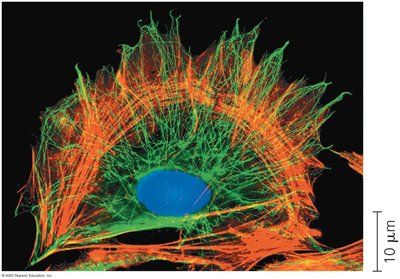
Support and Shape: The cytoskeleton maintains the cell's architecture, preventing it from collapsing or deforming.
Motility: It enables movement of the cell as a whole (e.g., amoeboid movement) and movement of organelles within the cell.
Organization: The cytoskeleton organizes the cell's interior, positioning organelles and facilitating intracellular transport.
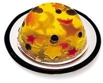
Key Point: The cell is not a simple, unstructured mass (like a Jell-O mold with organelles); it is a highly organized system maintained by the cytoskeleton.
Cytoskeletal Components
Microtubules
Microtubules are the largest cytoskeletal fibers, composed of tubulin protein subunits. They serve as tracks for organelle movement, are essential for chromosome separation during cell division, and power the movement of cilia and flagella.
Structure: Hollow tubes made of α- and β-tubulin dimers.
Functions:
Guide vesicle and organelle transport via motor proteins (e.g., kinesin, dynein).
Form the mitotic spindle for chromosome movement during mitosis and meiosis.
Constitute the structural core of cilia and flagella, enabling their beating motion.
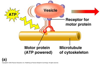
Example: Microtubules guide the movement of vesicles from the Golgi apparatus to the plasma membrane.
Centrosomes and Centrioles
The centrosome is the main microtubule-organizing center in animal cells. It contains a pair of centrioles, which are involved in organizing microtubules during cell division.
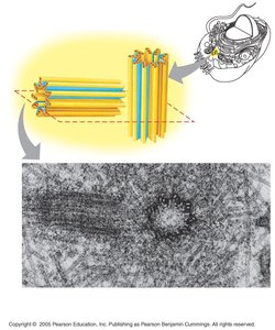
Centrosome: Region near the nucleus where microtubules are initiated.
Centrioles: Cylindrical structures composed of microtubule triplets, important for spindle formation.
Microfilaments (Actin Filaments)
Microfilaments are the thinnest cytoskeletal fibers, composed of actin protein subunits. They are crucial for cell shape, muscle contraction, cell movement, and cytoplasmic streaming.
Structure: Two intertwined strands of actin.
Functions:
Enable muscle contraction by interacting with myosin.
Drive amoeboid movement and the formation of pseudopodia.
Facilitate cytoplasmic streaming in plant cells, distributing nutrients and organelles.
Form the contractile ring during animal cell division (cytokinesis).
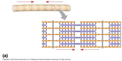
Example: Actin filaments enable white blood cells to move toward infection sites by changing cell shape.
Intermediate Filaments
Intermediate filaments are cytoskeletal fibers with a diameter between microtubules and microfilaments. They are composed of various proteins, such as keratins, and provide mechanical strength to cells.
Structure: Rope-like fibers made of different proteins (e.g., keratin, vimentin).
Functions:
Maintain cell shape and resist tension.
Anchor organelles, including the nucleus.
Form the nuclear lamina, supporting the nuclear envelope.
Example: Intermediate filaments in skin cells (keratin) provide resilience against mechanical stress.
Exterior Structures of Eukaryotic Cells
Plant Cell Walls
The cell wall is a rigid structure that surrounds plant cells, providing protection, maintaining cell shape, and preventing excessive water uptake. It is primarily composed of cellulose microfibrils embedded in a matrix of other polysaccharides and proteins.
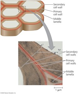
Primary Cell Wall: Thin and flexible, formed first as the cell grows.
Secondary Cell Wall: Thicker and stronger, deposited after cell growth stops.
Middle Lamella: Pectin-rich layer that glues adjacent cells together.
Example: The rigidity of plant stems and leaves is due to the presence of cell walls.
Extracellular Matrix (ECM) of Animal Cells
The extracellular matrix (ECM) is a complex network of proteins and carbohydrates outside animal cells. It provides structural support, mediates cell signaling, and helps cells adhere to one another.
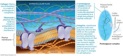
Main Components:
Collagen: Provides tensile strength.
Proteoglycans: Form a gel-like matrix for support and hydration.
Fibronectin: Connects ECM components to cell surface receptors (integrins).
Integrins: Transmembrane proteins that link the ECM to the cytoskeleton, transmitting signals between the cell's exterior and interior.
Example: The ECM is essential for tissue integrity and wound healing in animals.
Cell Junctions in Animal Tissues
Cell junctions are specialized structures that connect adjacent animal cells, facilitating communication and maintaining tissue integrity.
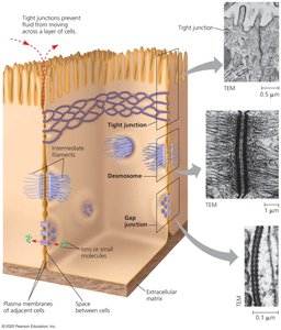
Tight Junctions: Seal neighboring cells together to prevent leakage of extracellular fluid.
Desmosomes: Anchor cells together, providing mechanical stability.
Gap Junctions: Allow ions and small molecules to pass directly between cells for communication.
Example: Gap junctions in cardiac muscle cells enable rapid transmission of electrical signals for synchronized contraction.
Plasmodesmata in Plant Cells
Plasmodesmata are channels that traverse plant cell walls, connecting the cytoplasm of adjacent cells and allowing the exchange of substances and signaling molecules.
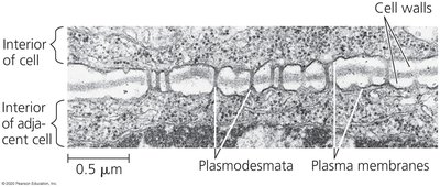
Function: Facilitate communication and transport of water, nutrients, and signaling molecules between plant cells.
Structure: Lined by plasma membrane, continuous with the endoplasmic reticulum of adjacent cells.
Example: Plasmodesmata enable the coordinated function of plant tissues by allowing the movement of hormones and other molecules.
