 Back
BackEukaryotic Chromosome Structure and Cell Division
Study Guide - Smart Notes
 Tailored notes based on your materials, expanded with key definitions, examples, and context.
Cell Division and Chromosome Structure
Overview of the Cell Cycle
The cell cycle is the series of events that cells undergo to grow and divide. It consists of several phases: G1 (first gap), S (DNA synthesis), G2 (second gap), and M (mitosis). During the S phase, DNA is replicated, preparing the cell for division.
G1 Phase: Cell grows and prepares for DNA replication.
S Phase: DNA is synthesized, resulting in duplicated chromosomes.
G2 Phase: Cell prepares for mitosis.
M Phase: Mitosis and cytokinesis occur, resulting in two daughter cells.
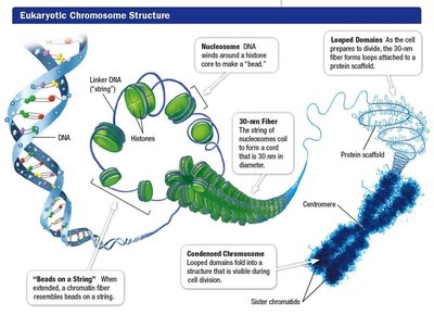
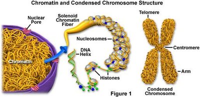
Eukaryotic Chromosome Structure
Eukaryotic chromosomes are highly organized structures composed of DNA and proteins. The DNA is wrapped around histone proteins, forming nucleosomes, which further coil and fold to create condensed chromosomes visible during cell division.
Chromatin: The complex of DNA and proteins that forms chromosomes.
Nucleosome: DNA wrapped around a histone core, forming the basic unit of chromatin.
30-nm Fiber: Nucleosomes coil into a thicker fiber, increasing compaction.
Condensed Chromosome: Chromatin further folds and loops, forming the highly condensed chromosome seen during mitosis.
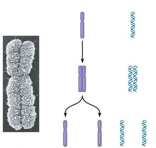
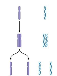
Chromosome Duplication and Sister Chromatids
Before cell division, each chromosome is duplicated, resulting in two identical sister chromatids joined at the centromere. During mitosis, these chromatids are separated and distributed to daughter cells.
Sister Chromatids: Identical copies of a chromosome, joined at the centromere.
Centromere: The region where sister chromatids are most tightly connected.
Cohesin: Protein complex that holds sister chromatids together.
Kinetochore: Protein structure at the centromere that attaches chromatids to spindle fibers.
Mitotic Spindle and Centrosomes
The mitotic spindle is essential for chromosome separation during mitosis. It is composed of microtubules that originate from centrosomes, which are microtubule-organizing centers in the cell.
Mitotic Spindle: Structure made of microtubules that guides chromosome movement.
Centrosome: Organelle that organizes microtubules and forms the spindle poles.
Microtubules: Protein filaments that make up the spindle and facilitate chromosome movement.

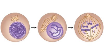
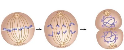
Phases of Mitosis
Mitosis is divided into several phases, each characterized by specific changes in chromosome and spindle structure:
Prophase: Chromosomes condense, spindle forms, nuclear envelope breaks down.
Metaphase: Chromosomes align at the cell equator (metaphase plate).
Anaphase: Sister chromatids separate and move to opposite poles.
Telophase: Chromosomes decondense, nuclear envelope reforms.
Cytokinesis: Cytoplasm divides, forming two daughter cells.
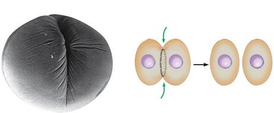
Cytokinesis in Animal Cells
Cytokinesis is the process by which the cytoplasm divides, resulting in two separate cells. In animal cells, this occurs through the formation of a cleavage furrow, which is created by a contracting ring of microfilaments.
Cleavage Furrow: Indentation that forms as the cell membrane is pinched inward.
Microfilaments: Actin filaments that contract to deepen the furrow.
Myosin: Motor protein that interacts with actin to facilitate contraction.
Summary Table: Key Structures in Chromosome Organization and Cell Division
Structure | Function |
|---|---|
Chromatin | DNA-protein complex; forms chromosomes |
Nucleosome | Basic unit of chromatin; DNA wrapped around histones |
Centromere | Joins sister chromatids; attachment site for kinetochore |
Cohesin | Protein complex holding sister chromatids together |
Kinetochore | Protein structure for spindle attachment |
Mitotic Spindle | Microtubule structure guiding chromosome movement |
Centrosome | Microtubule organizing center |
Cleavage Furrow | Site of cytokinesis in animal cells |
Additional info:
Chromatin can exist in a less condensed (euchromatin) or highly condensed (heterochromatin) state, affecting gene expression.
Telomeres are specialized regions at chromosome ends, protecting DNA from degradation.
