 Back
BackExam 4 Study Guide: Osmoregulation, Circulation, Nerves, Muscles, and Hormones
Study Guide - Smart Notes
 Tailored notes based on your materials, expanded with key definitions, examples, and context.
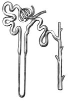
Q6. Label the nephron.
Background
Topic: Structure and function of the nephron in the kidney
This question tests your understanding of the anatomy of the nephron, which is the functional unit of the kidney responsible for filtration, reabsorption, secretion, and excretion.
Key Terms:
Bowman's capsule
Glomerulus
Proximal tubule
Loop of Henle (descending and ascending limbs)
Distal tubule
Collecting duct
Step-by-Step Guidance
Start by identifying the glomerulus, which is a cluster of capillaries where blood filtration begins.
Locate Bowman's capsule, which surrounds the glomerulus and collects the filtrate.
Trace the filtrate as it moves into the proximal tubule, where reabsorption of water, ions, and nutrients occurs.
Follow the path into the loop of Henle, noting the descending limb (permeable to water) and ascending limb (permeable to salts).
Continue to the distal tubule, which further adjusts the composition of the filtrate.
Try solving on your own before revealing the answer!
Final Answer:
The nephron includes the following labeled parts: glomerulus, Bowman's capsule, proximal tubule, descending limb of loop of Henle, ascending limb of loop of Henle, distal tubule, and collecting duct.
Each part plays a specific role in the filtration and processing of blood to form urine.
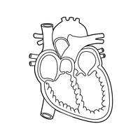
Q15. Label all the components from above (except the capillary beds) in the heart diagram.
Background
Topic: Anatomy of the heart and major blood vessels
This question tests your ability to identify the main chambers, valves, and vessels of the heart, which are essential for understanding circulation.
Key Terms:
Atrium (right and left)
Ventricle (right and left)
Aorta
Vena cava
Pulmonary artery and vein
Semilunar valve
Atrioventricular valve
Step-by-Step Guidance
Identify the four chambers: right atrium, right ventricle, left atrium, and left ventricle.
Locate the major blood vessels: aorta, vena cava, pulmonary artery, and pulmonary vein.
Find the valves: semilunar valves (between ventricles and arteries) and atrioventricular valves (between atria and ventricles).
Label each structure on the diagram, making sure to distinguish between oxygenated and deoxygenated blood flow.
Try solving on your own before revealing the answer!
Final Answer:
The heart diagram should be labeled with the right and left atria, right and left ventricles, aorta, vena cava, pulmonary artery, pulmonary vein, semilunar valves, and atrioventricular valves.
These components are critical for understanding the flow of blood through the heart and the circulatory system.
