 Back
BackFlow of Genetic Information: DNA Replication, Transcription, and Translation
Study Guide - Smart Notes
 Tailored notes based on your materials, expanded with key definitions, examples, and context.
Introduction to the Flow of Genetic Information
The flow of genetic information in all cells, both prokaryotic and eukaryotic, follows a central dogma: DNA is replicated before cell division, transcribed into RNA, and then translated into proteins. These processes are fundamental to cell biology and are mediated by a variety of enzymes and molecular machines. Understanding these pathways is essential for comprehending how genetic information is maintained and expressed in living organisms.
Nucleic Acids: Structure and Function
DNA and RNA Structure
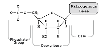
Nucleic acids are polymers composed of nucleotide monomers. The two main types are deoxyribonucleic acid (DNA) and ribonucleic acid (RNA). Each nucleotide consists of three components: a phosphate group, a pentose sugar, and a nitrogenous base. DNA contains the sugar deoxyribose and the bases adenine (A), thymine (T), cytosine (C), and guanine (G). RNA contains ribose and the bases A, C, G, and uracil (U) instead of thymine. Nucleotides are linked by phosphodiester bonds, giving the molecule directionality from the 5’ to 3’ end.
Base Pairing and Double Helix
DNA is double-stranded, with complementary base pairing: A pairs with T (or U in RNA), and C pairs with G. The two strands are antiparallel, forming a double helix. RNA is typically single-stranded.
Proteins: Structure and Directionality
Proteins are polymers of amino acids, linked by peptide bonds. Each amino acid has a central carbon, an amine group, a carboxyl group, and a variable R group. Proteins have directionality, with an N-terminus (free amine) and a C-terminus (free carboxyl). After synthesis, polypeptides fold into functional three-dimensional structures.
DNA Replication
Overview of DNA Replication
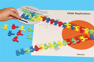
DNA replication is the process by which a cell copies its DNA before division. In eukaryotes, this occurs in the nucleus and begins at multiple origins of replication (ORI). The enzyme helicase unwinds the double helix, creating replication bubbles with two replication forks at each end. Single-strand binding proteins stabilize the unwound DNA.
Enzymes and Steps in Replication
Primase synthesizes short RNA primers complementary to the template DNA.
DNA polymerase III extends the primers, synthesizing new DNA in the 5’ to 3’ direction.
The leading strand is synthesized continuously toward the replication fork.
The lagging strand is synthesized discontinuously, forming Okazaki fragments away from the fork.
DNA polymerase I replaces RNA primers with DNA.
Ligase seals gaps between fragments, completing the sugar-phosphate backbone.
Multiple replication bubbles allow rapid duplication of large genomes, such as the human genome.
Transcription: DNA to RNA
Mechanism of Transcription
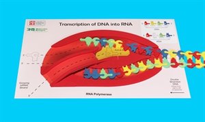
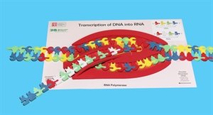
Transcription is the synthesis of RNA from a DNA template. It begins at a promoter region, where RNA polymerase binds and unwinds the DNA. Only one DNA strand serves as the template. RNA polymerase adds complementary RNA nucleotides in the 5’ to 3’ direction until it reaches a terminator sequence.

mRNA (messenger RNA): Encodes protein sequences.
rRNA (ribosomal RNA) and tRNA (transfer RNA): Noncoding RNAs essential for translation.
Translation: RNA to Protein
The Genetic Code and Codons
Translation converts the nucleotide sequence of mRNA into a polypeptide chain. The genetic code is read in triplets called codons, each specifying an amino acid. There are 64 possible codons but only 20 amino acids, so the code is degenerate (some amino acids are specified by more than one codon).
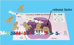
Ribosomes and tRNA
Ribosomes are complexes of rRNA and protein, with three sites: A (aminoacyl), P (peptidyl), and E (exit). tRNAs carry specific amino acids and have anticodons that pair with mRNA codons. Translation begins at the start codon (AUG, coding for methionine) and ends at a stop codon (UAA, UAG, UGA).
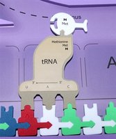
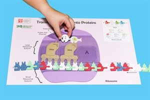
Initiation: Small ribosomal subunit binds mRNA; initiator tRNA binds start codon; large subunit assembles.
Elongation: Aminoacyl tRNAs enter the A site; peptide bonds form; ribosome translocates along mRNA.
Termination: Stop codon is reached; release factor binds; polypeptide is released.
Summary Table: Key Differences Between DNA and RNA
Feature | DNA | RNA |
|---|---|---|
Sugar | Deoxyribose | Ribose |
Bases | A, T, C, G | A, U, C, G |
Strandedness | Double-stranded | Single-stranded |
Function | Genetic storage | Information transfer, catalysis |
Key Terms and Concepts
Phosphodiester bond: Covalent bond linking nucleotides in a strand.
Hydrogen bond: Weak bond between complementary bases in DNA.
Antiparallel: Orientation of DNA strands in opposite directions.
Okazaki fragments: Short DNA fragments synthesized on the lagging strand.
Codon: Three-nucleotide sequence in mRNA specifying an amino acid.
Anticodon: Three-nucleotide sequence in tRNA complementary to mRNA codon.
Release factor: Protein that terminates translation at stop codons.
Equations and Formulas
General structure of a nucleotide:
Directionality:
Base pairing:
Applications and Importance
Understanding the flow of genetic information is crucial for fields such as genetics, molecular biology, biotechnology, and medicine. Errors in replication, transcription, or translation can lead to mutations and disease. Techniques such as PCR, DNA sequencing, and gene editing rely on these fundamental processes.
