 Back
BackLESSON 4: Form and Function of Cells and Tissues: Cytoskeleton, Intercellular Connections, and Basic Tissue Types
Study Guide - Smart Notes
 Tailored notes based on your materials, expanded with key definitions, examples, and context.
Form and Function of Cells and Tissues
Introduction
This study guide covers the structural and functional organization of cells and tissues, focusing on the cytoskeleton, intercellular junctions, and the four basic tissue types. Understanding these concepts is essential for grasping how cells maintain their shape, interact with each other, and organize into tissues that perform specialized functions in multicellular organisms.
Cytoskeleton: Structure and Function
Overview of the Cytoskeleton
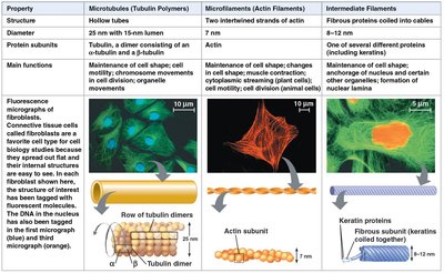
The cytoskeleton is a dynamic network of protein filaments that provides structural support, determines cell shape, and facilitates intracellular transport and cellular movement. It consists of three main components: microtubules, microfilaments (actin filaments), and intermediate filaments.
Microtubules: Hollow tubes made of tubulin; involved in maintaining cell shape, moving organelles, and chromosome separation during cell division.
Microfilaments (Actin Filaments): Thin strands of actin; support cell shape, enable movement, and form the contractile ring during cytokinesis.
Intermediate Filaments: Rope-like fibers made of various proteins (e.g., keratin); provide mechanical strength and anchor organelles.
Microtubules
Microtubules are the largest cytoskeletal filaments and play a key role in intracellular transport and cell division.
Composed of α- and β-tubulin dimers.
Form tracks for motor proteins (e.g., kinesin, dynein) to transport vesicles and organelles.
Essential for the formation of the mitotic spindle during cell division.

Microfilaments (Actin Filaments)
Microfilaments are the thinnest cytoskeletal elements and are concentrated beneath the plasma membrane.
Composed of actin monomers.
Support cell shape, especially at the cell cortex.
Enable cell movement (e.g., muscle contraction, cell crawling) in conjunction with myosin.
Form microvilli in epithelial cells to increase surface area for absorption.

Intermediate Filaments
Intermediate filaments provide tensile strength and help cells withstand mechanical stress.
Composed of various proteins, such as keratins in epithelial cells.
Stabilize cell structure and anchor organelles.
Form the nuclear lamina, supporting the nuclear envelope.
Summary Table: Cytoskeletal Elements
Cell Surface Protrusions: Microvilli, Cilia, and Flagella
Microvilli
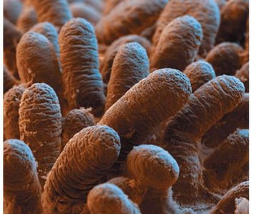
Microvilli are finger-like extensions of the plasma membrane supported by actin filaments. They increase the surface area for absorption, especially in intestinal epithelial cells.
Cilia and Flagella
Cilia and flagella are motile structures composed of microtubules arranged in a 9+2 pattern. Cilia are usually numerous and move substances across the cell surface, while flagella are longer and typically move the cell itself (e.g., sperm cells).
Cilia: Short, many per cell, move fluid or substances (e.g., respiratory tract).
Flagella: Long, usually one per cell, move the cell (e.g., sperm).
Comparison of Cilia and Flagella
Feature | Cilia | Flagella |
|---|---|---|
Number per cell | Many | One or few |
Length | Short | Long |
Function | Move substances relative to cell | Move the cell itself |
Intercellular Connections
Types of Intercellular Junctions
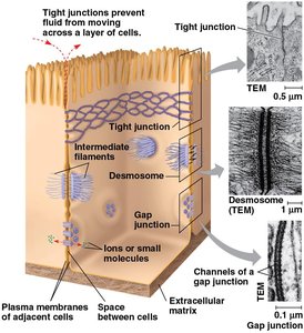
Cells in tissues are connected by specialized junctions that provide structural integrity and communication.
Tight Junctions: Seal adjacent cells to prevent leakage of extracellular fluid.
Desmosomes: Anchor cells together, providing mechanical strength.
Gap Junctions: Allow direct communication between cells via channels for ions and small molecules.
Basic Tissue Types
Overview of Tissue Organization
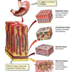
Multicellular organisms are organized into four basic tissue types, each with specialized functions. Most organs contain all four tissue types.
Epithelial Tissue: Covers body surfaces and lines cavities.
Connective Tissue: Supports, binds, and protects organs.
Muscle Tissue: Enables movement through contraction.
Nervous Tissue: Receives, processes, and transmits information.
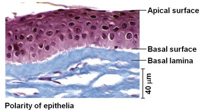
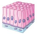
Epithelial Tissue
Epithelial tissue forms protective barriers and is specialized for absorption, secretion, and sensation. It exhibits polarity, with an apical (external) and basal (internal) surface.
Simple Squamous Epithelium: Flat cells for diffusion (e.g., air sacs in lungs).
Stratified Squamous Epithelium: Multiple layers for protection (e.g., skin).
Simple Columnar Epithelium: Tall cells for absorption/secretion (e.g., intestines).
Pseudostratified Columnar Epithelium: Appears layered, often ciliated (e.g., respiratory tract).
Connective Tissue
Connective tissue supports and binds other tissues. It consists of cells scattered within an extracellular matrix (ECM) composed of protein fibers and ground substance.
Loose Connective Tissue: Binds epithelia to underlying tissues; contains fibrocytes.
Fibrous Connective Tissue: Dense collagen fibers; found in tendons and ligaments.
Bone: Mineralized matrix; cells are osteocytes.
Adipose Tissue: Stores fat; cells are adipocytes.
Cartilage: Flexible matrix; cells are chondrocytes.
Blood: Liquid ECM (plasma); cells include erythrocytes, leukocytes, and thrombocytes.
Muscle Tissue
Muscle tissue is specialized for contraction and movement, powered by actin and myosin filaments.
Skeletal Muscle: Voluntary, striated, multinucleated; moves the skeleton.
Smooth Muscle: Involuntary, non-striated; found in walls of organs.
Cardiac Muscle: Striated, found in the heart; cells connected by intercalated discs for synchronized contraction.
Nervous Tissue
Nervous tissue is responsible for receiving, processing, and transmitting information. It consists of neurons and supporting glial cells.
Neurons: Transmit electrical signals; composed of cell body, axon, and dendrites.
Glial Cells: Support, nourish, and insulate neurons.
Microscopy in Cell and Tissue Study
Types of Microscopy
Microscopy is essential for visualizing cells and tissues. The main types include:
Light Microscopy (LM): Uses visible light; includes brightfield, phase contrast, and fluorescence microscopy.
Electron Microscopy (EM): Uses electron beams; includes scanning (SEM) and transmission (TEM) electron microscopy.
Key Concepts in Microscopy
Magnification: Ratio of image size to actual size.
Resolution: Minimum distance between two distinguishable points.
Contrast: Difference in visual properties that makes objects distinguishable.
Steps for Using a Light Microscope
Place the slide on the stage.
Turn the nosepiece to the 10x objective.
Find the image with the coarse focus knob.
Turn the nosepiece to the 20x objective.
Correctly set the diaphragm and condenser.
Adjust the aperture diaphragm.
Focus with the fine focus knob.
Summary Table: Four Basic Tissue Types
Tissue Type | Main Function | Location/Example |
|---|---|---|
Epithelial | Lining, secretion, absorption | Skin, lining of gut |
Connective | Support, strength, elasticity | Tendons, bone, blood |
Muscle | Movement | Skeletal muscles, heart |
Nervous | Information processing, communication | Brain, nerves |
Additional info: The cytoskeleton is not only structural but also highly dynamic, allowing cells to rapidly reorganize in response to environmental cues. The extracellular matrix (ECM) in connective tissue is crucial for tissue integrity and signaling. Understanding microscopy is foundational for all cell and tissue studies in biology.
