 Back
BackFrom Cells to Organ Systems: Structure and Function of Tissues
Study Guide - Smart Notes
 Tailored notes based on your materials, expanded with key definitions, examples, and context.
Tissues: Groups of Cells with a Common Function
Definition and Overview
Tissues are groups of specialized cells that are similar in structure and perform a common function. Multiple tissue types may combine to form organs, which carry out specific functions in the body. There are four primary tissue types in humans:
Epithelial tissues
Connective tissues
Muscle tissues
Nervous tissues
Cell Junctions
Cell junctions are specialized structures that hold adjacent cells together and facilitate communication or movement.
Tight junctions: Seal plasma membranes tightly together, preventing leakage (e.g., digestive tract lining, bladder lining).
Adhesion junctions (spot desmosomes): Permit some movement between cells, allowing tissues to stretch and bend (e.g., skin).
Gap junctions: Protein channels enable movement of materials between adjacent cells (e.g., liver, heart).
Epithelial Tissues
Structure and Function
Epithelial tissues are organized as sheets of cells, one or more layers thick. Their main purposes are to line body cavities, cover surfaces, and protect underlying tissues.
Glandular epithelia: Epithelial cells adapted to form glands.
Glands: Specialized epithelial tissues that synthesize and secrete products.
Exocrine glands: Secrete products into hollow organs or ducts.
Endocrine glands: Secrete hormones into the blood for distribution throughout the body.
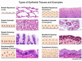
Classification by Shape
Epithelial tissues are classified based on cell shape:
Squamous: Flattened cells; form outer surface of skin, line blood vessels, lungs, mouth, throat, vagina.
Cuboidal: Cube-shaped cells; form lining of kidney tubules, glandular tissue.
Columnar: Tall, rectangular cells; line digestive tract, certain reproductive organs, larynx. May include goblet cells that secrete mucus.
Classification by Number of Layers
Simple: Single-layered; adapted for diffusion across cell barriers. Found in glands, respiratory, digestive, and reproductive systems.
Stratified: Multiple-layered; provide protection, as in the skin surface.
Distribution in the Human Body
Different types of epithelial tissues are distributed throughout the body, each serving specific functions in organs and systems. 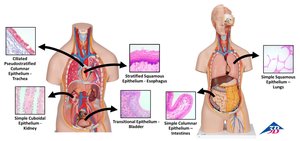
The Basement Membrane
The basement membrane is a noncellular layer directly beneath epithelial tissue, composed of proteins secreted by epithelial cells and connective tissue. It provides structural support and attaches the epithelial layer to underlying connective tissue. 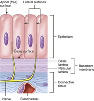
Connective Tissue
General Functions and Structure
Connective tissue supports and connects body parts, stores fat, and produces blood cells. It consists of three main components:
Ground substance: Aqueous solution containing water, proteins, and polysaccharides.
Fibers: Collagen (strong, slightly flexible), elastic (stretchable), and reticular (thin, interconnecting).
Cells: Fibroblasts, macrophages, lymphocytes, neutrophils, fat cells, mast cells, white blood cells.
Types of Connective Tissue Proper
Loose connective tissues: Areolar, adipose, reticular.
Dense connective tissues: Dense regular, dense irregular, elastic.
Specialized Connective Tissues
Cartilage: Transitional tissue from which bone develops; maintains shape of nose and ears; cushions vertebrae; lines joint cavities. Structure: dense connective tissue of collagen fibers, ground substance produced by chondroblasts, mature cells called chondrocytes. Slow to heal due to lack of blood vessels.
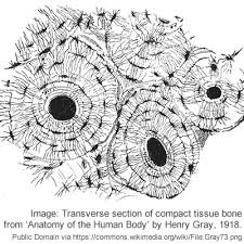
Bone: Forms the skeleton; contains few living cells; matrix composed of calcium phosphate; contains numerous blood vessels.
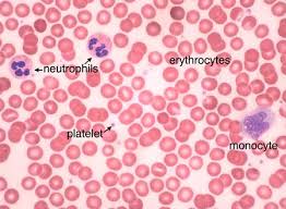
Blood: Fluid matrix (plasma) with red blood cells (transport oxygen), white blood cells (immune defense), and platelets (blood clotting).
Muscle Tissue
Structure and Function
Muscle tissue contracts to produce movement. Muscle cells (muscle fibers) are long, thin, and arranged parallel to each other. There are three types:
Skeletal muscle: Moves body parts; voluntary; activated by nerves; fibers have multiple nuclei.
Cardiac muscle: Located only in the heart; involuntary; cells are shorter, blunt-ended, one nucleus per cell; gap junctions enable coordinated contraction.
Smooth muscle: Surrounds hollow organs and tubes (blood vessels, digestive tract, uterus, bladder); involuntary; slim, tapered cells with one nucleus; gap junctions enable coordinated contraction.
Nervous Tissue
Structure and Function
Nervous tissue forms a rapid communication network including the brain, spinal cord, and nerves.
Neuron: Specialized cell that generates and transmits electrical impulses. Components: cell body (nucleus, cytoplasm), dendrites (receive signals), axon (transmits impulses).
Glial cells: Surround and protect neurons; provide nutrients.
Organs and Organ Systems
Definition and Examples
Organs are structures composed of two or more tissue types joined together to perform specific functions. Example: the heart, which pumps blood and contains cardiac muscle, smooth muscle, nervous tissue, connective tissue, and epithelial tissue.
Organ Systems
Organ systems are groups of organs that serve broad functions essential for survival. There are eleven organ systems in the human body, such as the digestive system (mouth, throat, stomach, intestines, liver, pancreas, gallbladder) and lymphatic system (defense against disease, circulation, transport of digestion products). 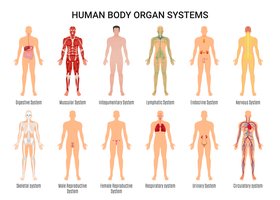
Homeostasis
Definition and Importance
Homeostasis is the relative constancy of conditions within the internal environment. Multicellular organisms devote significant metabolic activity to maintaining homeostasis.
Interstitial fluid: Clear fluid between cells; source of nutrients and repository for wastes.
Negative Feedback Control System
Negative feedback systems detect and counteract deviations from normal.
Controlled variable: Any property that must be regulated (e.g., body temperature).
Sensor: Monitors current value and sends information to control center.
Control center: Compares value to set point, signals effector if necessary.
Effector: Takes action to correct imbalance.
Example: Regulation of core body temperature by hypothalamus, sensors in skin and organs, effectors such as blood vessels, sweat glands, and skeletal muscles.
Positive Feedback
Positive feedback amplifies events and is relatively uncommon in living organisms. It is not a mechanism for maintaining homeostasis. Example: Childbirth.
The Skin: An Organ of the Integumentary System
Structure and Function
The integumentary system includes skin, hair, nails, and glands. Functions include protection from dehydration and injury, defense against microorganisms, regulation of body temperature, synthesis of vitamin D, and sensation.
Skin Layers
Epidermis: Outer layer; multiple layers of stratified squamous epithelial cells; no blood vessels; contains keratinocytes (produce keratin), basal cells (dividing keratinocytes), and melanocytes (produce melanin).
Dermis: Primarily dense connective tissue; contains collagen, elastic, and reticular fibers; fibroblasts, mast cells, white blood cells, fat cells; supports tissues and provides strength and elasticity.
Accessory Structures of Dermis
Hair: Shaft above skin, root below surface in follicle.
Smooth muscle: Raises hair to upright position.
Sebaceous (oil) glands: Secrete sebum to moisten and soften skin.
Sweat glands: Secrete sweat for temperature regulation; contains antimicrobial peptides.
Blood vessels: Supply nutrients, remove waste, assist in temperature regulation.
Sensory nerve endings: Detect heat, cold, touch, pressure, vibration.
Vitamin D synthesis: Cholesterol-like molecule in skin converted to inactive vitamin D, activated by liver and kidneys.
